
A patient sits down and says a specialist told them they have “Ménière’s.” The first honest question a doctor can ask back is whether it’s a disease or a syndrome — and the Ménière’s disease vs syndrome distinction is not pedantry, because the word you choose quietly admits how much of this condition medicine still cannot explain. This article draws a clear line between what is settled, what is still argued over, how the condition is diagnosed and named, how it relates to sudden low-frequency hearing loss, and how it is treated when an attack hits versus during the quiet stretches in between.
Ménière’s Disease vs Ménière’s Syndrome: Which Term Is Correct?
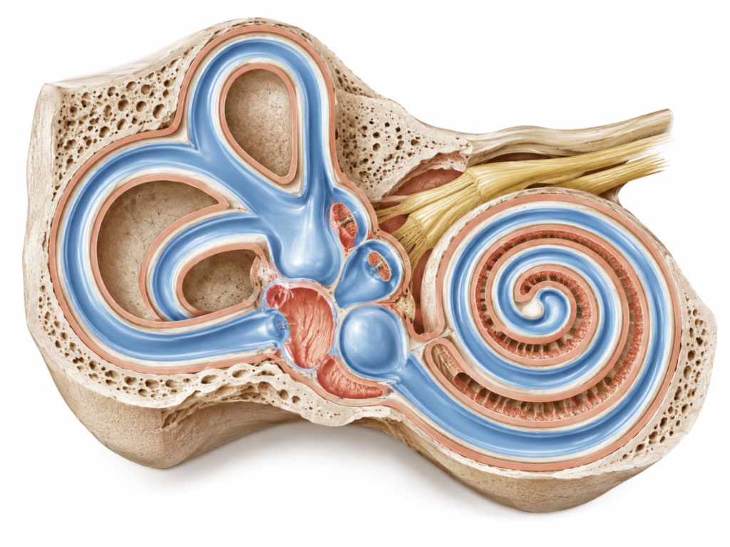
Both terms are correct — but for different situations. “Ménière’s disease” is the precise term when the cause is unknown, which describes the large majority of patients. “Ménière’s syndrome” is reserved for the same cluster of symptoms when a specific underlying cause has been identified — for example head trauma, autoimmune inner-ear disease, an acoustic neuroma, or superior semicircular canal dehiscence.
The cleanest way to think about it is the Bell’s palsy rule. Once you find the cause of a facial paralysis, it stops being “Bell’s palsy” and becomes paralysis due to that cause. In the same way, once you identify why someone’s inner ear is generating vertigo, it stops being Ménière’s disease and becomes Ménière’s syndrome secondary to that cause.
For everyday clinical purposes, the international consensus criteria standardise on the term “Ménière’s disease,” applied only after other explanations have been excluded [Lopez-Escamez, Diagnostic criteria for Ménière’s disease, 2015]. So for the typical patient whose cause is never found, “Ménière’s disease” is the technically accurate label. In casual conversation the two words get used interchangeably, but that loose usage hides a real point: calling it a “disease” is a way of saying we don’t yet know why this is happening.
What We Know vs What We’re Still Guessing
The most useful thing a patient can be given is an honest boundary between established fact and open question.
| ✅ Established | ❓ Still hypothesis / debated |
|---|---|
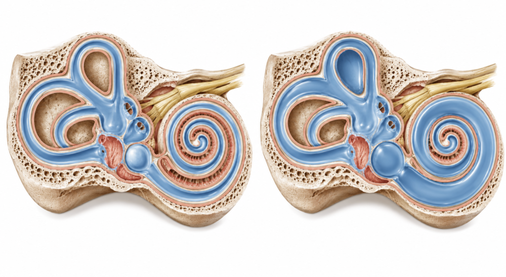
| The pathological hallmark is endolymphatic hydrops — a swelling of the inner ear’s endolymph compartment | Whether that swelling causes the symptoms, or is a byproduct of the real underlying process |
| Temporal-bone studies find hydrops in nearly every Ménière’s ear examined | The initiating cause of the hydrops itself, which is unknown by definition |
| The clinical picture is a recognisable set: episodic vertigo, fluctuating low-to-mid-frequency hearing loss, tinnitus, and ear fullness | The trigger of a single acute attack — several competing theories exist, none proven |
| Hydrops can now be seen in living patients on specialised gadolinium-enhanced MRI | Why hydrops shows up in some people with no symptoms at all |
| The condition behaves as multifactorial, with genetic, immune, and vascular contributions | Whether it is truly one disease or a family of look-alike conditions |
The imaging point is worth dwelling on. For more than seventy years, hydrops could only be confirmed at autopsy, but high-resolution MRI now lets clinicians visualise it in a living, symptomatic patient [Gürkov, What is Menière’s disease? A contemporary re-evaluation of endolymphatic hydrops, 2016]. That advance has sharpened the debate rather than ending it — because seeing the swelling still doesn’t prove the swelling is what generates the vertigo.
This is the single most misunderstood point about the condition. Patient leaflets, health websites, and even some clinical summaries describe the fluid swelling as the cause of Ménière’s — full stop. That overstates the evidence. Hydrops is the most consistent finding in the disease, but “the most consistent finding” and “the proven cause” are not the same claim, and the space between them is exactly what the next two sections are about.
Clinical Perspective: Hydrops is the fingerprint at the scene. A fingerprint tells you someone was there — it does not, by itself, tell you what they did.
Why the Cause Is Still Debated
If the swelling is always there, why is anyone still arguing? Because the correlation is not as clean as it first looks, and reasonable experts read the same evidence differently.
The tidy explanation — many possible causes lead to hydrops, and hydrops in turn produces the symptoms — has a name in the literature: the “final common pathway” hypothesis. For it to be true, every case of Ménière’s should show hydrops, and every case of hydrops should produce Ménière’s symptoms. The first half holds up. The second half does not.
In a landmark review of human temporal bones, every case with classic symptoms showed hydrops in at least one ear, yet a substantial number of hydropic ears came from people who never had the classic symptoms in life [Merchant, Pathophysiology of Meniere’s syndrome: are symptoms caused by endolymphatic hydrops?, 2005]. On that basis, the authors argued hydrops should be treated as a histologic marker of the condition rather than the direct cause of the symptoms.
A later meta-analysis of 541 hydropic temporal bones read the data the other way. It concluded the autopsy evidence does not support hydrops being a mere epiphenomenon, leaving causation as the most likely interpretation — but with a crucial caveat: hydrops alone is insufficient, so one or more cofactors (the authors flagged vascular risk factors) must convert silent hydrops into symptomatic disease [Foster, Endolymphatic hydrops in Ménière’s disease: cause, consequence, or epiphenomenon?, 2013]. Same bones, opposite emphasis. Add the absence of any gold-standard diagnostic test and a presentation that varies wildly between patients, and you have a condition whose science has stayed unsettled for decades.
The Best-Accepted Explanation Today
Here is the current consensus, stated plainly: endolymphatic hydrops is the central mechanism, but it is “necessary but not sufficient.” Hydrops is present in nearly every affected ear, yet swelling on its own does not produce the disease. As the autopsy meta-analysis concluded, if hydrops is causative, it is insufficient by itself, and one or more cofactors must convert silent hydrops into symptomatic disease [Foster, Endolymphatic hydrops in Ménière’s disease: cause, consequence, or epiphenomenon?, 2013]. That is why the condition is now framed as multifactorial — hydrops arising in an ear that also carries additional vulnerabilities, most clearly vascular risk factors, with immune and genetic susceptibility also implicated — rather than a single fluid problem.
So the honest headline is not “excess fluid causes vertigo.” It is closer to: disordered inner-ear fluid regulation produces hydrops, and in a susceptible ear, additional hits turn that silent hydrops into episodic disease.
Clinical Perspective: Hydrops sets the stage. Something else has to pull the trigger — and that “something else” probably differs from one patient to the next, which is part of why a treatment that works beautifully for one person does little for another.
Symptoms: Definite vs Probable Ménière’s
Clinically, Ménière’s announces itself through four core symptoms: rotational vertigo attacks, fluctuating low-to-mid-frequency sensorineural hearing loss, tinnitus, and a sense of fullness or pressure in the affected ear. The international criteria then sort patients into two diagnostic buckets — definite and probable — which differ in specific, testable ways [Lopez-Escamez, Diagnostic criteria for Ménière’s disease, 2015].
| Definite Ménière’s | Probable Ménière’s | |
|---|---|---|
| Vertigo episodes | Two or more spontaneous episodes, each lasting 20 minutes to 12 hours | Two or more episodes of vertigo or dizziness, each lasting 20 minutes to 24 hours (wider window) |
| Hearing loss | Audiometrically documented low-to-mid-frequency loss in the affected ear (before, during, or after an attack) | No audiometry requirement |
| Aural symptoms | Fluctuating hearing, tinnitus, or fullness in the affected ear | Fluctuating hearing, tinnitus, or fullness in the affected ear |
| Exclusion | Not better explained by another vestibular disorder | Not better explained by another vestibular disorder |
Read the table and the difference becomes concrete. What separates “probable” from “definite” is essentially two things: probable does not require hearing loss proven on an audiogram, and it allows attacks to run longer, up to 24 hours [Webster, Systemic pharmacological interventions for Ménière’s disease, 2023]. A patient with textbook spinning attacks and ear fullness but a stubbornly normal audiogram on the day of testing may sit in the “probable” category until a hearing change is finally captured.
One rule cuts across both categories: the diagnosis is only valid after ruling out mimics, and the most important mimic is vestibular migraine, which the guideline explicitly directs clinicians to assess for [Basura, Clinical Practice Guideline: Ménière’s Disease, 2020]. Overlapping symptoms make the two easy to confuse.
Acute Low-Tone Hearing Loss vs Ménière’s — and Can It Progress?
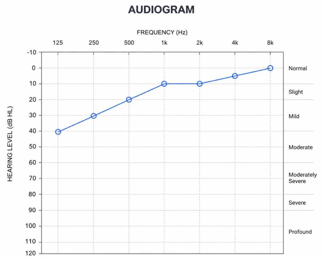
Some patients don’t arrive with vertigo at all. They arrive with a sudden, isolated drop in the low frequencies — a muffled ear, a low-pitched roar, a feeling of fullness — and normal hearing higher up. This is acute low-tone sensorineural hearing loss (ALHL), and it sits close enough to Ménière’s to raise an obvious worry: is this the start of it?
The two conditions are biologically related. Recent inner-ear MRI work in patients with ALHL who had no vertigo still found measurable cochlear hydrops on the affected side — concentrated at the apex of the cochlea, exactly where low-frequency sound is processed [Seo, Findings of intravenous gadolinium inner ear magnetic resonance imaging in patients with acute low-tone sensorineural hearing loss, 2023]. In other words, ALHL appears to reflect hydrops largely confined to the cochlea, whereas full Ménière’s also drags in the balance organs and produces vertigo. The overlap is real, but so is the distinction.
So can ALHL turn into Ménière’s? Yes — but only in a minority. In that same MRI cohort, hearing improved in most patients, recurrence was common at around 47%, and roughly 16% progressed to Ménière’s disease over follow-up [Seo, Findings of intravenous gadolinium inner ear magnetic resonance imaging in patients with acute low-tone sensorineural hearing loss, 2023]. A broad scoping review of 41 studies reached the same shape of answer: most patients recover their hearing, recurrence and fluctuation are frequent, and progression to Ménière’s happens in only a minority [Huang, Prognosis of acute low-tone hearing loss without vertigo: a scoping review, 2023].
The practical takeaway for a worried patient is reassuring but not dismissive. Isolated low-tone hearing loss is not Ménière’s disease. But low-tone loss that arrives together with true spinning vertigo, ear fullness, and tinnitus deserves closer follow-up, because that combination is the profile more likely to declare itself as Ménière’s over time.
Treatment: Acute Attacks vs Maintenance
Treatment splits cleanly into two jobs that are easy to conflate: stopping the misery of an attack in progress, and reducing how often attacks happen in the first place. They call for different tools.
During an acute attack, the goal is symptom control. Clinicians typically reach for vestibular suppressants and anti-nausea medication to blunt the vertigo and vomiting, alongside rest and hydration until the storm passes. Notably, the guideline specifically advises against using vestibular rehabilitation to manage an acute vertigo attack — rehabilitation has its place, but not in the middle of a crisis [Basura, Clinical Practice Guideline: Ménière’s Disease, 2020].
Between attacks, prevention follows a stepwise ladder, escalating only if simpler measures fail and weighing every step against the patient’s remaining hearing.
| Step | Approach | Notes |
|---|---|---|
| 1 — Conservative | Low-sodium diet, trigger and lifestyle management, betahistine, diuretics | First-line and low-risk; betahistine is widely used, though the trial evidence supporting it is weak |
| 2 — Non-ablative | Intratympanic corticosteroid injections | Aims to control attacks while preserving hearing |
| 3 — Ablative | Intratympanic gentamicin | Effective for vertigo but carries a real risk of hearing loss; reserved for active disease failing gentler therapy |
| 4 — Surgical / last resort | Endolymphatic sac surgery, or labyrinthectomy when hearing is already non-usable | For disabling, treatment-resistant disease |
A dose of honesty belongs on that first rung. When the available randomised trials for common preventive drugs were pooled, the evidence was rated low or very low certainty, meaning we have limited confidence that the reported effects reflect the true benefit [Webster, Systemic pharmacological interventions for Ménière’s disease, 2023]. These treatments are used because they are reasonable and often helpful in practice, not because the science behind them is airtight.
Two supports run alongside the ladder regardless of step: vestibular rehabilitation for the chronic imbalance that can linger between attacks, and hearing aids or assistive technology for the hearing loss itself [Basura, Clinical Practice Guideline: Ménière’s Disease, 2020].
Clinical Perspective: The natural history offers real reassurance. In long-term data pooling thousands of patients, the vertigo attacks that frighten people most tend to diminish within five to ten years, while most of the hearing decline also occurs in that same early window before settling [Huppert, Long-term course of Menière’s disease revisited, 2010]. There is no cure yet, but for most people this becomes a managed condition, not a life sentence of spinning.
Key Takeaways
- “Ménière’s disease” means the condition with no identifiable cause; “Ménière’s syndrome” means the same symptoms with a known underlying cause.
- The one thing we know is that nearly every Ménière’s ear shows endolymphatic hydrops — what we don’t know is whether that swelling is the cause or a byproduct.
- The current consensus is that hydrops is necessary but not sufficient, and Ménière’s is best understood as multifactorial.
- Definite Ménière’s requires audiogram-proven low-to-mid-frequency hearing loss; probable does not, and allows attacks lasting up to 24 hours.
- Acute low-tone hearing loss (ALHL) can progress to Ménière’s, but only in a minority — around one in six in recent data — with higher risk when vertigo and fullness accompany it.
- Treatment is stepwise, from diet and lifestyle to intratympanic injections to surgery; there is no cure, but symptoms are manageable and attacks often lessen over time.
Frequently Asked Questions
Is it Ménière’s disease or Ménière’s syndrome? It is a disease when no cause can be found, which is most cases, and a syndrome when a specific cause is identified. The distinction mirrors Bell’s palsy: once the underlying cause of the inner-ear symptoms is known, the label shifts from “disease” to “syndrome secondary to” that cause. International diagnostic criteria use “Ménière’s disease” as the standard term after other explanations have been excluded.
What actually causes Ménière’s disease? The honest answer is that we don’t fully know. The pathological hallmark is endolymphatic hydrops, a swelling of the inner ear’s fluid compartment, but hydrops alone doesn’t reliably produce symptoms. The prevailing view is that hydrops must combine with additional genetic, immune, or vascular factors to become symptomatic disease, which is why it’s now considered multifactorial rather than a single fluid problem.
Can low-tone hearing loss turn into Ménière’s disease? Sometimes, but only in a minority of patients. Recent studies of acute low-tone hearing loss found that most people recover their hearing, recurrence is common, and roughly one in six go on to develop Ménière’s disease. The risk is higher when the hearing loss arrives together with vertigo, tinnitus, and ear fullness rather than in isolation.
What’s the difference between definite and probable Ménière’s? Definite Ménière’s requires audiogram-documented low-to-mid-frequency hearing loss and vertigo attacks lasting 20 minutes to 12 hours. Probable Ménière’s requires neither audiometric proof of hearing loss nor that shorter duration limit, allowing attacks of up to 24 hours. Both require that the symptoms not be better explained by another disorder, especially vestibular migraine.
How do you stop a Ménière’s attack? An acute attack is managed with vestibular-suppressant and anti-nausea medication, plus rest and hydration until it passes. This is different from long-term prevention, which follows a separate stepwise plan. Vestibular rehabilitation is specifically not recommended for an attack in progress, though it helps with chronic imbalance between attacks.
Is Ménière’s disease curable? No cure currently exists, but the condition is manageable. A stepwise treatment ladder — from diet and lifestyle changes to intratympanic injections and, rarely, surgery — controls vertigo in most patients. Reassuringly, attacks often become less frequent over the years, even as hearing tends to decline gradually.
References
- Lopez-Escamez JA, Carey J, Chung WH, Goebel JA, Magnusson M, Mandalà M, Newman-Toker DE, Strupp M, Suzuki M, Trabalzini F, Bisdorff A. Diagnostic criteria for Menière’s disease. J Vestib Res. 2015;25(1):1-7.
- Basura GJ, Adams ME, Monfared A, Schwartz SR, Antonelli PJ, Burkard R, et al. Clinical practice guideline: Ménière’s disease executive summary. Otolaryngol Head Neck Surg. 2020;162(4):415-434.
- Merchant SN, Adams JC, Nadol JB Jr. Pathophysiology of Meniere’s syndrome: are symptoms caused by endolymphatic hydrops? Otol Neurotol. 2005;26(1):74-81.
- Foster CA, Breeze RE. Endolymphatic hydrops in Ménière’s disease: cause, consequence, or epiphenomenon? Otol Neurotol. 2013;34(7):1210-1214.
- Gürkov R, Pyykö I, Zou J, Kentala E. What is Menière’s disease? A contemporary re-evaluation of endolymphatic hydrops. J Neurol. 2016;263 Suppl 1:S71-S81.
- Webster KE, Galbraith K, Harrington-Benton NA, Judd O, Kaski D, Maarsingh OR, MacKeith S, Ray J, Van Vugt VA, Burton MJ. Systemic pharmacological interventions for Ménière’s disease. Cochrane Database Syst Rev. 2023;2(2):CD015171.
- Seo HW, Kim Y, Kim HJ, Chung WH, Cho YS. Findings of intravenous gadolinium inner ear magnetic resonance imaging in patients with acute low-tone sensorineural hearing loss. Clin Exp Otorhinolaryngol. 2023;16(4):334-341.
- Huang RJ, Del Risco A, Riska KM, Cooper MW, Clark NW, Kaplan SJ, Kaylie D, Francis HW. Prognosis of acute low-tone hearing loss without vertigo: a scoping review. Laryngoscope. 2023;133(10):2457-2469.
- Huppert D, Strupp M, Brandt T. Long-term course of Menière’s disease revisited. Acta Otolaryngol. 2010;130(6):644-651.
Joonpyo Hong, MD is a board-certified otolaryngologist practicing in Korea. This article reflects his clinical interpretation of published research and does not constitute individual medical advice.
For more interesting content:
https://curiousmd.com/vestibular-neuritis/
https://curiousmd.com/perilymphatic-fistula-from-weightlifting/
Linkt out to: