
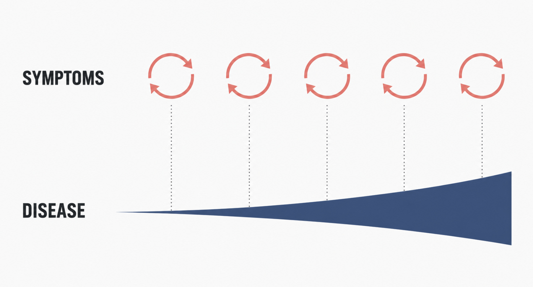
Cholesteatoma symptoms are easy to dismiss. A small amount of foul-smelling discharge from one ear, recurring three or four times over two years, clearing with antibiotic drops each time. No pain. Under the microscope, the only abnormal finding may be a tiny crust in the upper portion of the eardrum — easy to mistake for wax.
That crust is often the visible tip of a cholesteatoma. By the time the disease announces itself more loudly, it has usually been dissolving bone for years.
This article covers what a cholesteatoma is, why it forms, how it is diagnosed, and — the question patients ask most — why the answer is surgery rather than medication. It deals with cholesteatoma of the middle ear, behind the eardrum. Cholesteatoma arising in the ear canal itself is a different disease with different management, and is covered separately.
What Is a Cholesteatoma? (Not a Tumor, But It Acts Like One)
A cholesteatoma is a cyst of keratin-producing squamous epithelium — ordinary skin — trapped inside the middle ear or mastoid, where skin does not belong. The name comes from the pearly sheen of the mass on cut section. It has nothing to do with cholesterol, and it is not a neoplasm.
Two layers matter, and the distinction drives everything that follows:
- The matrix — living epithelium that continuously produces keratin.
- The keratin debris — the accumulated product, filling the sac.
Skin normally sheds its keratin outward. Inside a closed middle ear space, there is no outward, and no route by which accumulated keratin can leave. The sac fills, expands, and presses against whatever surrounds it. The lesion is benign histologically and destructive behaviorally, and the mismatch between those two facts is the most common reason patients underestimate it.
What Causes Cholesteatoma?
The retraction pocket route (most common)
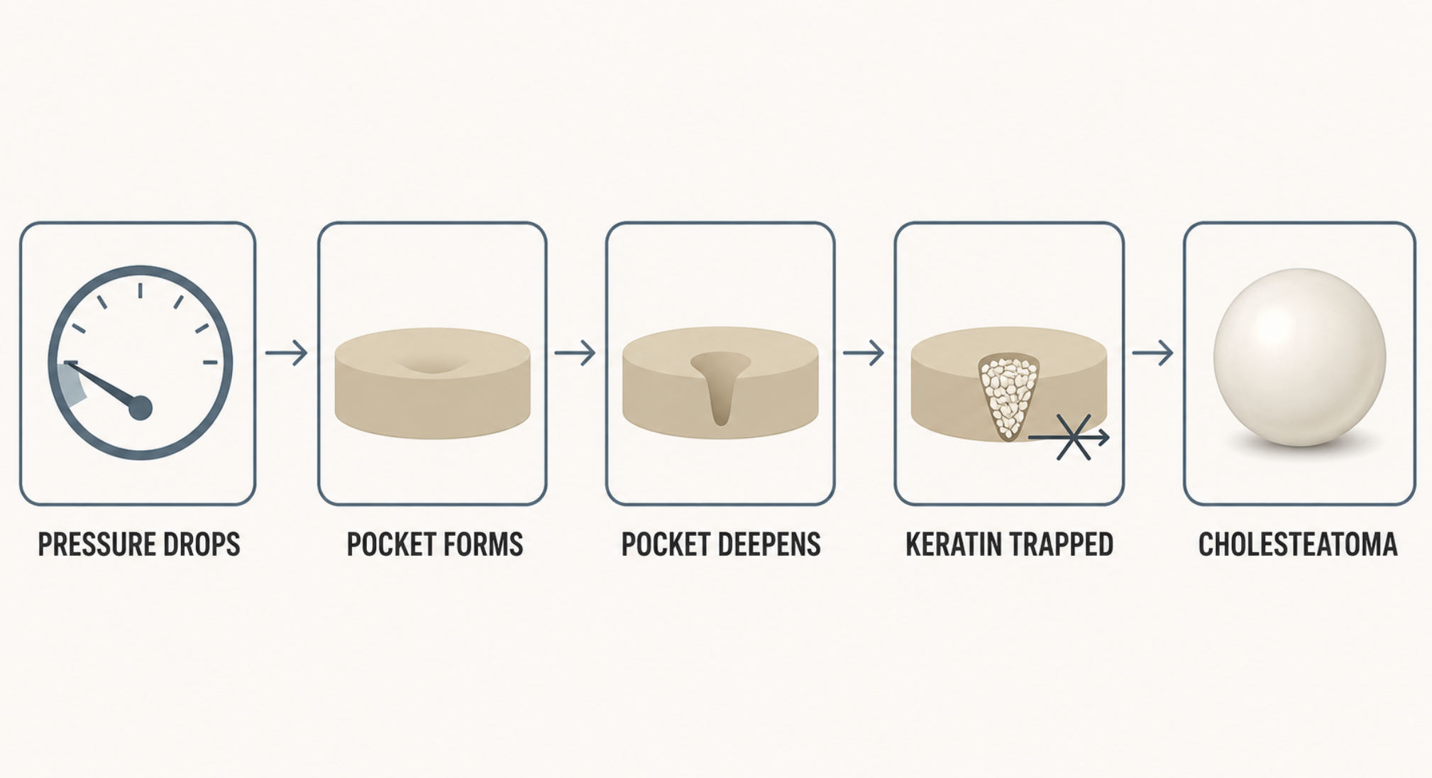
The usual sequence begins with eustachian tube dysfunction. The eustachian tube equalizes pressure between the middle ear and the back of the nose; when it fails chronically, negative pressure develops behind the eardrum. The weakest part of the drum — usually the pars flaccida in the attic — is drawn inward, forming a retraction pocket.
A shallow pocket cleans itself. A deep one cannot. Keratin accumulates in the recess, the pocket deepens under continued negative pressure, and at some point self-cleaning fails permanently. That is the moment a retraction pocket becomes a cholesteatoma [Kuo, Updates and Knowledge Gaps in Cholesteatoma Research, 2015].
Through a perforation
Skin from the ear canal can also migrate into the middle ear through an existing hole in the eardrum, whether from chronic infection, trauma, or previous surgery. This is the secondary acquired form.
Congenital cholesteatoma
A small subset arises behind an entirely intact, normal-looking eardrum, with no history of ear infection, perforation, or ear surgery. These are typically found in children as an incidental white mass during an examination for something else.
Who is at higher risk
Children carry more risk than adults: a shorter, more horizontal eustachian tube, and more frequent upper respiratory infections. A history of recurrent otitis media, ventilation tubes, chronic allergic rhinitis, adenoid hypertrophy, or cleft palate all raise the probability. Most cases are acquired rather than congenital [Kuo, A Review of Current Progress in Acquired Cholesteatoma Management, 2015].
Early Cholesteatoma Symptoms — and Why They Get Missed
Early cholesteatoma is quiet. The classic picture is:
- Scant, foul-smelling discharge from one ear, recurring over months or years
- Discharge that improves with antibiotic drops and then returns
- Gradual conductive hearing loss on that side
- A sensation of fullness
- In advanced disease: vertigo, facial weakness, headache
Notice what is missing. There is usually no pain. Painlessness gets read as reassurance, which inverts its actual meaning — in cholesteatoma, pain tends to arrive when a complication does.
The second trap is the antibiotic response. Drops treat the bacteria colonizing the sac, so the smell and drainage stop. Nothing has happened to the sac.
Clinical Perspective
Late diagnosis usually follows not from a missed examination but from a series of successful ones. Several visits, each ending with drops that worked, each reasonable on its own terms. One-sided, recurrent, painless otorrhea warrants examination under the microscope before it receives a third prescription.
How Is Cholesteatoma Diagnosed?
Diagnosis is clinical first, radiologic second. Each test answers a different question, and confusing them is a common source of both over- and under-treatment.
| Test | The question it answers |
|---|---|
| Otomicroscopy / endoscopy | Is there a lesion, and can the depth of the pocket be seen? |
| Audiogram and tympanometry | Has hearing or the ossicular chain already been damaged? |
| High-resolution temporal bone CT | How far has it spread? Is there erosion of the ossicles, facial canal, lateral semicircular canal, or tegmen? |
| Non-EPI diffusion-weighted MRI | Is this soft tissue actually cholesteatoma, or granulation and scar? |
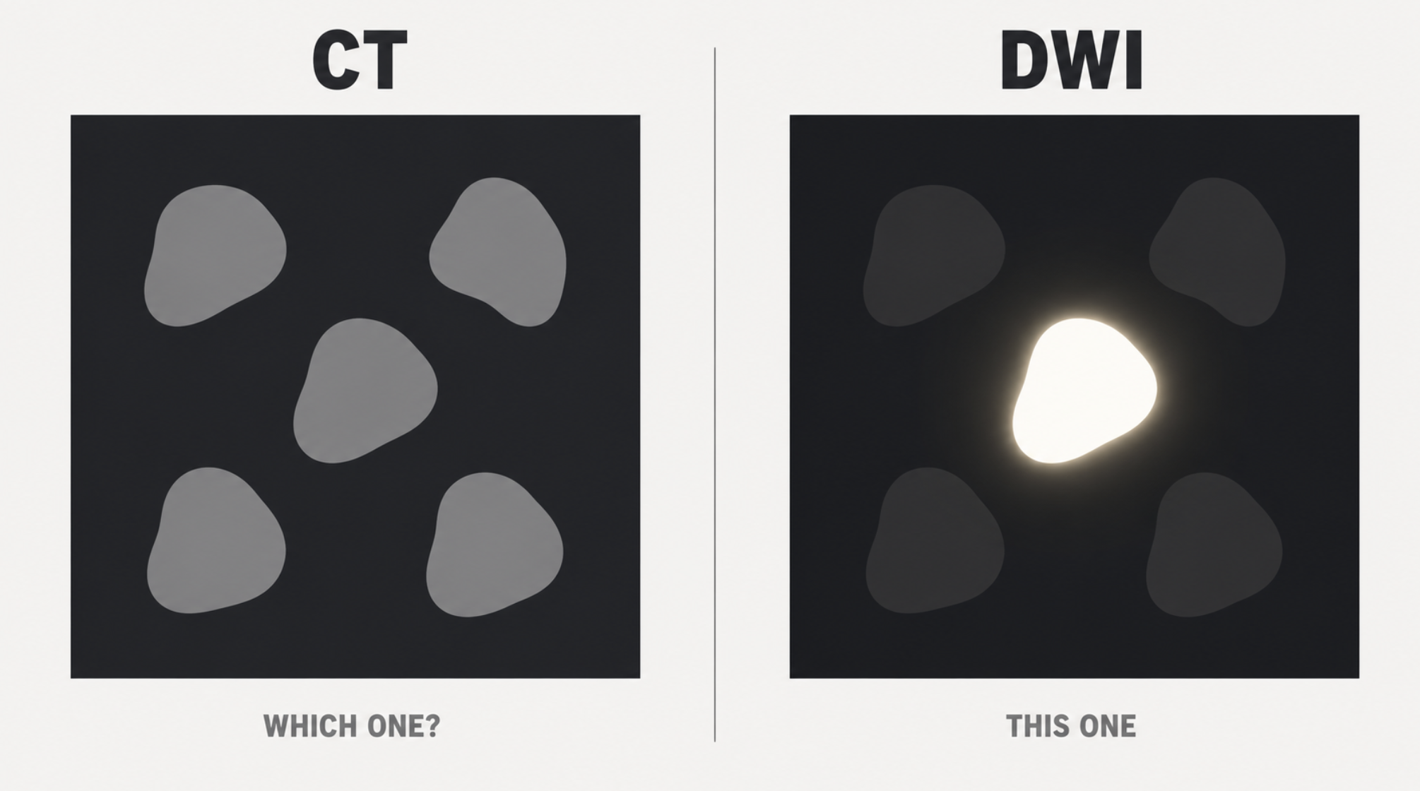
CT is excellent at showing bone and poor at characterizing soft tissue. It will show a shadow filling the attic; it cannot reliably distinguish keratin from granulation tissue or postoperative fibrosis.
Diffusion-weighted MRI answers that question directly. Keratin restricts water diffusion, producing a bright signal that granulation tissue does not. Non-echo-planar sequences avoid the susceptibility artifact that degrades echo-planar imaging at the skull base, and can resolve lesions of only a few millimeters [De Foer, Detection of Postoperative Residual Cholesteatoma with Non-Echo-Planar Diffusion-Weighted Magnetic Resonance Imaging, 2008]. A meta-analysis restricted to studies with surgical correlation found pooled sensitivity of 91% and specificity of 92% for non-echo-planar DWI in detecting middle ear cholesteatoma [Lingam, A Meta-Analysis on the Diagnostic Performance of Non-Echoplanar Diffusion-Weighted Imaging in Detecting Middle Ear Cholesteatoma, 2017], and systematic review confirms non-EPI sequences outperform EPI for residual and recurrent disease [Muzaffar, Diffusion-Weighted Magnetic Resonance Imaging for Residual and Recurrent Cholesteatoma, 2017].
The practical consequence is significant: a negative scan can spare a patient a second operation performed solely to look.
Staging: why not every cholesteatoma operation is the same
Two patients with the same diagnosis can be offered visibly different operations, and the reason is anatomical extent. The European Academy of Otology and Neurotology and the Japanese Otological Society published a joint consensus on definitions, classification, and staging of middle ear cholesteatoma, dividing the middle ear into four regions — supratubal recess, tympanic cavity including the sinus tympani, attic, and mastoid — and assigning stages by how many are involved and whether complications are present [Yung, EAONO/JOS Joint Consensus Statements on the Definitions, Classification and Staging of Middle Ear Cholesteatoma, 2017].
A lesion confined to the attic and a lesion filling the mastoid with a tegmen defect are the same disease at different stages, and they call for different operations.
Why Surgery Is the Definitive Treatment
Keratin does not dissolve, and the middle ear cannot clear it
No enzyme, drop, or antibiotic breaks down accumulated keratin, and a closed sac offers no path for it to drain. Whatever is produced stays.
The matrix is living skin — leave it, and it rebuilds
Suctioning debris from a pocket in clinic empties it temporarily. If the matrix remains, it refills. The surgical objective is therefore not evacuation of debris but complete removal of the epithelium producing it — which is also why partial or piecemeal removal is a poor bargain.
Antibiotics treat the infection, not the disease
Cholesteatoma sacs become secondarily colonized, and that colonization is what causes the smell and drainage. Topical antibiotics address it effectively while the sac continues to expand underneath. A symptom-free interval after treatment is therefore weak evidence about the disease itself.
Expansion is what erodes bone
A mechanically expanding sac combined with chronic inflammation at its margin resorbs surrounding bone. Which structures are threatened depends largely on where the sac sits. Attic disease characteristically involves the long process of the incus early; more extensive disease reaches the lateral semicircular canal, the fallopian canal carrying the facial nerve, and the tegmen separating the middle ear from the middle cranial fossa [Kuo, Updates and Knowledge Gaps in Cholesteatoma Research, 2015].
What definitive treatment actually means
Complete surgical excision under general anesthesia is the standard of care. The reason is not surgical preference but exposure: adequate visualization of the attic, sinus tympani, and mastoid is what allows the matrix to be removed in full, and residual matrix is the single most avoidable cause of recurrent disease. The only genuine exception is a patient who cannot safely undergo anesthesia.
One situation demonstrates the logic better than any statistic. When the opposite ear has no useful hearing, operating on the only hearing ear is a frightening proposition — yet removal usually remains the preferred option, because the disease poses a greater long-term threat to that hearing than a well-planned operation does.
What Happens If You Wait?
Untreated disease progresses along a fairly consistent sequence:
- Conductive hearing loss from ossicular erosion
- Chronic otorrhea and recurrent infection
- Labyrinthine fistula, producing vertigo — most often at the lateral semicircular canal
- Facial nerve palsy where the fallopian canal is eroded or dehiscent
- Sensorineural hearing loss once the inner ear is involved, which is not reversible
- Intracranial complications — meningitis, brain abscess, sigmoid sinus thrombosis
The last category deserves context rather than alarm. Serious intracranial complications are the least common items on this list, and how often they appear depends heavily on the health system a patient lives in — access to specialist care, availability of antibiotics, and how early the disease is caught. Where those conditions are favorable, most cholesteatomas are found and treated long before the tegmen is at risk. The argument for operating is not that catastrophe is likely. It is that timely surgery removes this entire list from the table.
What the Surgery Involves
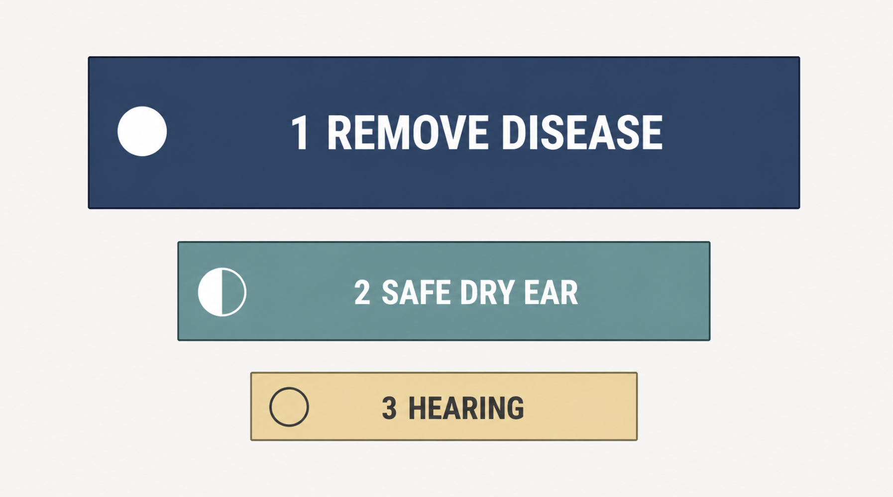
The goals, in priority order: complete removal of the disease, a safe and dry ear, and hearing reconstruction. Hearing is genuinely third, and setting that expectation beforehand prevents a great deal of postoperative disappointment.
Canal wall up versus canal wall down. The canal wall up approach preserves the bony ear canal and normal anatomy; canal wall down removes it, creating an open mastoid cavity with better long-term disease control. Meta-analysis found a substantially higher rate of postoperative disease with the intact canal wall approach compared with canal wall down [Tomlin, Surgical Technique and Recurrence in Cholesteatoma, 2013], and a later systematic review in adults reached the same conclusion [Kerckhoffs, The Disease Recurrence Rate After the Canal Wall Up or Canal Wall Down Technique in Adults, 2016]. The tradeoff runs in the other direction too: an open cavity requires periodic cleaning, sometimes indefinitely, and imposes water restrictions and some other uncomfortable situations compare to canal wall up approach.
Endoscopic ear surgery. For disease confined to the middle ear, the endoscope reaches around corners a microscope cannot — the sinus tympani and anterior attic in particular — and can avoid a mastoidectomy altogether in selected cases.
Reconstruction. Tympanoplasty repairs the drum; ossiculoplasty restores the sound conduction chain using titanium prostheses or autologous cartilage. Reconstruction is sometimes deferred to a second stage to confirm the disease is gone first, which means hearing may be temporarily worse after the first operation than before it.
After Surgery: Recurrence and Follow-Up
Two distinct things can happen, and the terminology matters because the implications differ. Residual disease is regrowth from matrix left behind. Recurrence is a new cholesteatoma forming from a new retraction pocket. The first reflects the completeness of the excision; the second reflects the eustachian tube problem that started the process, which does not disappear because the ear was operated on.
Historically every canal wall up patient returned for a second-look operation. Increasingly, non-EPI DWI surveillance substitutes for that, reserving reoperation for patients with a positive scan [van Egmond, A Systematic Review of Non-Echo Planar Diffusion-Weighted Magnetic Resonance Imaging for Detection of Primary and Postoperative Cholesteatoma, 2016].
Imaging has limits worth stating plainly. False negatives cluster in small and thin, plaque-like lesions; wax in the canal can produce false positives. A negative scan reduces the probability of residual disease considerably but does not replace continued clinical examination.
When It’s Not the Middle Ear
Not all keratin in an ear is middle ear cholesteatoma. Impacted wax, keratosis obturans, and cholesteatoma arising in the bony ear canal itself can all present as white debris filling the canal — and debris from an attic cholesteatoma can spill outward and look exactly like a canal problem. That overlap is precisely why microscopic examination, rather than a glance with an otoscope, is the diagnostic starting point. These canal conditions should be covered in a separate article.
Clinical Perspective
The most common question about cholesteatoma is whether medication can be tried first.
The most useful answer is two sentences. Keratin does not dissolve. The skin producing it is alive, and if it stays, the sac refills. Everything else about the recommendation follows from those two facts.
What patients find harder to accept is that the operation is not merely the best available option but the only one that ends the process — and that its success depends on something they cannot see, namely whether the surgeon could reach every recess where matrix might hide. That is the real reason this is a general anesthesia procedure rather than an office one. Adequate exposure is not a matter of comfort. It is what separates a cure from a postponement.
Key Takeaways
- Cholesteatoma is not cancer, but it enlarges, erodes bone, and does not resolve on its own.
- The classic early presentation is painless, foul-smelling, one-sided ear discharge that keeps returning — which is why it is so often mistaken for wax.
- Antibiotics and ear drops treat the infection colonizing a cholesteatoma; they do not remove the lesion, and bone erosion continues while symptoms improve.
- Surgery is necessary because keratin does not dissolve and the epithelium producing it will refill any sac left behind; complete excision requires the exposure that general anesthesia allows.
- CT defines the extent of disease; non-echo-planar diffusion-weighted MRI determines whether soft tissue is cholesteatoma or scar, and can replace second-look surgery in selected patients.
FAQ
Can a cholesteatoma go away on its own?
No. Keratin does not dissolve, and a closed middle ear sac has no drainage route, so the lesion enlarges over time rather than resolving. Periods of improvement reflect control of secondary infection, not regression of the cholesteatoma.
Can antibiotics or ear drops cure a cholesteatoma?
No. Drops treat the bacteria colonizing the sac, which stops the discharge and the smell. The sac itself is unaffected and continues to expand, so bone erosion can progress during a symptom-free interval.
Is cholesteatoma cancer?
No. It is a benign lesion made of ordinary skin in the wrong location. It is treated with the seriousness of a tumor because it grows and destroys bone locally, not because it is malignant or can spread elsewhere in the body.
Is surgery always necessary?
Yes, with one exception. Complete excision under general anesthesia is the standard treatment, because removing the matrix in full requires exposure that office procedures cannot provide. The only genuine exception is a patient who cannot safely undergo anesthesia.
What happens if a cholesteatoma is left untreated?
It progresses from conductive hearing loss to vertigo from labyrinthine fistula, facial nerve weakness, irreversible sensorineural hearing loss, and — uncommonly — intracranial complications such as meningitis or brain abscess. Timely surgery removes this progression.
How is cholesteatoma diagnosed?
Microscopic ear examination comes first. An audiogram documents functional damage, temporal bone CT maps the extent and any bone erosion, and non-echo-planar diffusion-weighted MRI distinguishes cholesteatoma from granulation tissue or scar when the CT finding is ambiguous.
Will my hearing come back after cholesteatoma surgery?
Sometimes, but it is not the primary goal. The first objective is complete removal of disease. When ossicular reconstruction is deferred to a second operation, hearing may be temporarily worse immediately after the first one.
Does cholesteatoma come back after surgery?
It can, in two forms — residual disease from matrix left behind and true recurrence from a new retraction pocket. Rates are higher in children and after canal wall up surgery, which is why long-term examination and imaging follow-up are standard.
References
- Yung M, Tono T, Olszewska E, Yamamoto Y, Sudhoff H, Sakagami M, et al. EAONO/JOS joint consensus statements on the definitions, classification and staging of middle ear cholesteatoma. J Int Adv Otol. 2017;13(1):1-8.
- Kuo CL, Shiao AS, Yung M, Sakagami M, Sudhoff H, Wang CH, et al. Updates and knowledge gaps in cholesteatoma research. Biomed Res Int. 2015;2015:854024.
- Kuo CL, Liao WH, Shiao AS. A review of current progress in acquired cholesteatoma management. Eur Arch Otorhinolaryngol. 2015;272(12):3601-3609.
- Lingam RK, Bassett P. A meta-analysis on the diagnostic performance of non-echoplanar diffusion-weighted imaging in detecting middle ear cholesteatoma: 10 years on. Otol Neurotol. 2017;38(4):521-528.
- Muzaffar J, Metcalfe C, Colley S, Coulson C. Diffusion-weighted magnetic resonance imaging for residual and recurrent cholesteatoma: a systematic review and meta-analysis. Clin Otolaryngol. 2017;42(3):536-543.
- van Egmond SL, Stegeman I, Grolman W, Aarts MC. A systematic review of non-echo planar diffusion-weighted magnetic resonance imaging for detection of primary and postoperative cholesteatoma. Otolaryngol Head Neck Surg. 2016;154(2):233-240.
- De Foer B, Vercruysse JP, Bernaerts A, Deckers F, Pouillon M, Somers T, et al. Detection of postoperative residual cholesteatoma with non-echo-planar diffusion-weighted magnetic resonance imaging. Otol Neurotol. 2008;29(4):513-517.
- Tomlin J, Chang D, McCutcheon B, Harris J. Surgical technique and recurrence in cholesteatoma: a meta-analysis. Audiol Neurootol. 2013;18(3):135-142.
- Kerckhoffs KG, Kommer MB, van Strien TH, Visscher SJ, Bruijnzeel H, Smit AL, et al. The disease recurrence rate after the canal wall up or canal wall down technique in adults. Laryngoscope. 2016;126(4):980-987.
Joonpyo Hong, MD is a board-certified otolaryngologist practicing in Korea. This article reflects his clinical interpretation of published research and does not constitute individual medical advice.
For more interesting content:
https://curiousmd.com/otomycosis/
https://curiousmd.com/age-related-hearing-loss-hear-not-understand/
https://curiousmd.com/cotton-swabs-in-your-ears/
Link out to:
- Cholesteatoma — American Academy of Otolaryngology–Head and Neck Surgery (ENT Health)
- Surgery for cholesteatoma — Healthdirect Australia
- Non-Echo-Planar Diffusion-Weighted MR Imaging for Cholesteatomas in High-Risk Retraction Pockets — American Journal of Neuroradiology