
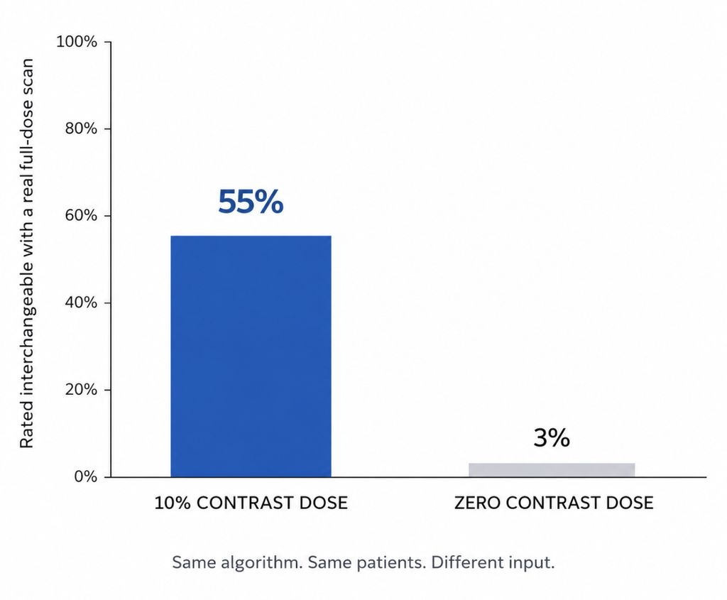
In a 2023 study, radiologists rated AI virtual contrast enhancement images as interchangeable with real full-dose scans in 55% of cases, when the algorithm was given a scan taken after 10% of a normal contrast dose. Given no contrast dose at all, that number fell to 3% [Haase R, Reduction of Gadolinium-Based Contrast Agents in MRI Using Convolutional Neural Networks and Different Input Protocols, 2023].
Those two numbers came from the same algorithm and the same patients. The only thing that changed was what went into the model. AI-generated contrast imaging is already running in clinics, though not in the way most people assume. What follows is what contrast injection actually buys a radiologist, what AI can and cannot replace today, and why the obstacle turns out not to be a shortage of training data.
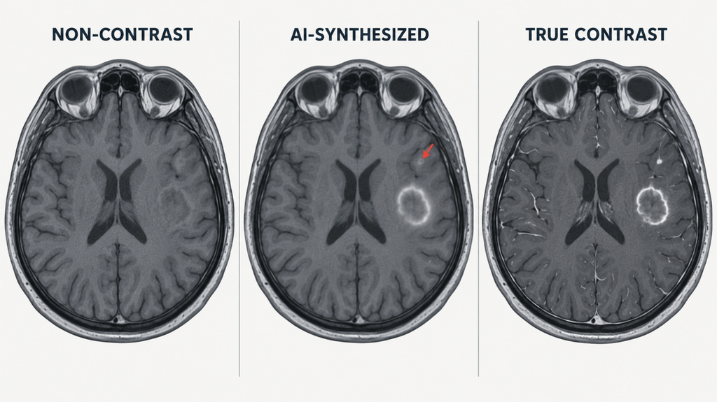
Why we inject contrast
Iodine for CT and gadolinium for MRI do one basic thing: they travel in blood and make blood-rich tissue brighter than everything around it.
That matters because so much of the body has near-identical density on a plain scan. A carotid artery, an adjacent lymph node, and a small tumor sitting between them can all look like the same shade of gray. Contrast separates them by circulation rather than by shape: how a tissue takes up blood, and how fast it lets it go.
Two settings benefit most. In cancer, tumors grow disorganized, leaky vessels that fill and drain differently from normal tissue, so contrast is what makes a lesion detectable, defines where its margin actually ends, and shows whether it is shrinking under treatment. In infection and inflammation, an abscess shows a bright rim around a dead center that stays dark, and that distinction decides whether a patient goes home on antibiotics or goes to the operating room.
| Non-contrast scan | With contrast | |
|---|---|---|
| Anatomy and size | Yes | Yes |
| Vessel vs. adjacent soft tissue | Often ambiguous | Separated |
| Tumor margin | Frequently indistinct | Defined |
| Abscess vs. solid mass | Hard to call | Rim enhancement pattern |
| Treatment response over time | Limited | Measurable |
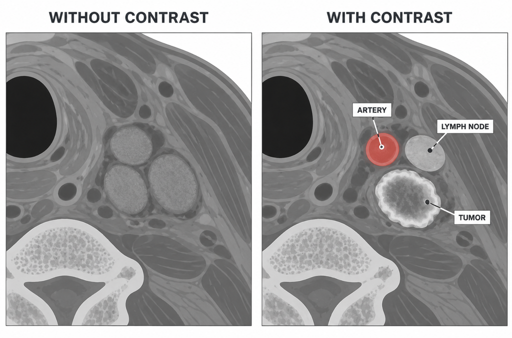
Why we’d rather skip it when we can
Contrast is safe for the overwhelming majority of patients. In a Korean multicenter registry of 196,081 patients, hypersensitivity reactions to iodinated contrast occurred in 0.73%, and severe reactions in 0.01% [Cha MJ, Hypersensitivity Reactions to Iodinated Contrast Media: A Multicenter Study of 196 081 Patients, 2019]. Those are small percentages of an enormous denominator.
Kidney concerns have been recalibrated rather than eliminated. The American College of Radiology and the National Kidney Foundation concluded that AKI risk after intravenous iodinated contrast in patients with reduced kidney function has been overstated, and drew a line between injury caused by contrast and injury merely coincident with it. They still recommend saline prophylaxis for patients below an eGFR of 30 who are not on maintenance dialysis [Davenport MS, Use of Intravenous Iodinated Contrast Media in Patients with Kidney Disease, 2020].
Gadolinium raises a separate issue, which is accumulation with repeated exposure. That falls hardest on patients scanned every few months for years: glioma follow-up, head and neck cancer surveillance.
None of this makes contrast dangerous. It makes the rule narrower. When contrast is needed, use it, and when the same information can be obtained without it, don’t. The second half of that rule is what AI has been aimed at.
What AI virtual contrast enhancement can do today
Two different problems tend to get discussed as one.
Amplification starts from a scan that already has some contrast in it. A 2018 Stanford study trained a network on pre-contrast plus 10%-dose images and recovered something close to full-dose quality [Gong E, Deep learning enables reduced gadolinium dose for contrast-enhanced brain MRI, 2018]. That line of work has since reached the clinic. A recent multi-center, multi-reader evaluation of an FDA-cleared contrast-boosting algorithm found improved lesion visualization and image quality without raising the administered dose, though the evaluation was conducted largely by the algorithm developer and a contrast manufacturer, which is worth weighing [Venkata SP, Deep-Learning Based Contrast Boosting: A Multi-Center Multi-Reader Study on Clinical Performance With Standard Contrast Enhanced Brain MRI, 2026]. This part is cleared and in use.
Zero-dose synthesis starts from nothing and tries to predict the contrast image from non-contrast sequences alone. The results there are mixed rather than uniformly negative.
An early feasibility study reached 91.8% sensitivity and 91.2% specificity for predicting enhancement from non-contrast brain MRI. But two radiologists in a Turing test correctly separated real from synthetic images 80% and 90% of the time, spotting blurred ring enhancement and omitted small vessels [Kleesiek J, Can Virtual Contrast Enhancement in Brain MRI Replace Gadolinium?: A Feasibility Study, 2019].
A large multicentre study went further and asked whether synthetic images were good enough for a specific clinical job, rather than good enough to fool a reader. Synthesized post-contrast sequences underestimated tumor volume slightly, but time to progression came out at 4.2 months with synthetic images versus 4.3 months with real ones, not a significant difference [Jayachandran Preetha C, Deep-learning-based synthesis of post-contrast T1-weighted MRI for tumour response assessment in neuro-oncology, 2021].
Progress varies by organ, and CT lags MRI substantially. Published CT work remains largely single-center and has not yet produced the multi-center reader studies that exist for MRI. In cardiac imaging, a model trained on 430 patients across three centers converted contrast-free cine images into scar maps with 91.27% sensitivity and 95.83% specificity [Qi H, Predicting Late Gadolinium Enhancement of Acute Myocardial Infarction in Contrast-Free Cardiac Cine MRI Using Deep Generative Learning, 2024]. In liver imaging, a diffusion-based protocol built from 1,769 patients matched conventional MRI for hepatocellular carcinoma detection while cutting acquisition from 28.1 to 4.1 minutes [Zhang Y, Deep learning empowered gadolinium-free contrast-enhanced abbreviated MRI for diagnosing hepatocellular carcinoma, 2025].
The real bottleneck
The obvious guess is that these models need more data, or that patient privacy is keeping the data locked away. The published results point elsewhere.
The 2023 Bonn study is the cleanest natural experiment available: same architecture, same patients, four different input settings. Interchangeability with the real full-dose scan was 55% with a 10% contrast dose and 3% with none. False positive findings averaged 0.42 per case in the low-dose setting and 2.00 per case in the zero-dose setting [Haase R, 2023]. The model did not degrade between those two runs. The information it needed had stopped being in the input.
Quantitative image quality and diagnostic usefulness also come apart. That same study reported excellent quantitative performance alongside limited interchangeability. A loss function averaged across an entire brain volume barely notices a 3 mm metastasis, because a handful of voxels contributes almost nothing to the score, even when those voxels are the entire reason the scan was ordered.
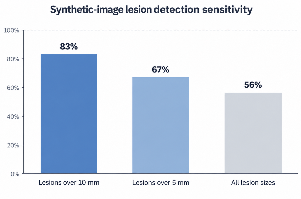
Lesion size is where the failure concentrates. A reader study using both pre-contrast and low-dose images as input, not zero dose, found synthetic images detected 83% of lesions larger than 10 mm, 67% of those larger than 5 mm, and 56% once all sizes were included. The authors concluded that full-dose injection remains essential for first-line diagnosis [Ammari S, Can Deep Learning Replace Gadolinium in Neuro-Oncology?: A Reader Study, 2022]. The shortfall appears even in the more favorable setting.
Generalization across hospitals is unresolved. A head and neck model reached an AUC of 0.95 on internal testing and 0.86 on an independent external set [Li Z, Deep learning-based non-contrast MRI model for nasopharyngeal carcinoma diagnosis, 2025]. The model was unchanged; only the hospital was.
Data scarcity explains part of this, but not the main part. What is actually scarce is paired studies with matched timing and scanner, along with coverage of rare pathology and post-surgical anatomy. Privacy restrictions are genuine, yet large public datasets exist and the failure modes above persist within them.
Validation is also circular. Amplification can be checked against the full-dose scan sitting right next to it. Zero-dose synthesis has to be validated against the very injection it aims to avoid, so building the evidence base requires doing the thing the method is meant to stop.
Clinical Perspective
Head and neck imaging is a good place to watch this play out. The nasopharynx is anatomically stable, and nasopharyngeal carcinoma patients get scanned repeatedly for years, so cumulative gadolinium exposure is a real clinical variable rather than a theoretical one.
The most notable recent result from that field took a different approach. Rather than synthesizing a contrast image for a radiologist to read, the model went directly from non-contrast MRI to a diagnosis, and outperformed virtual-contrast-enhancement baselines. In a 13-reader study, readers using non-contrast MRI with AI assistance reached an AUC of 0.90 against 0.93 for contrast-enhanced imaging [Li Z, 2025].
That changes the question. The useful goal may be working out which decisions actually require a contrast image, rather than teaching a model to draw one convincingly. Those are different engineering problems, and the second looks more tractable.
The published work also keeps pointing at one distinction: finding a lesion for the first time is not the same job as tracking one that is already known. Synthesis fails at detection and holds up in surveillance. A ninth follow-up scan in a patient whose tumor is mapped is a very different task from a first scan in a patient with a new symptom, and surveillance is the likelier place for contrast-free AI to establish itself first.
Key Takeaways
- AI-generated contrast imaging is already FDA-cleared and clinically deployed, but only for amplifying scans that already contain a reduced contrast dose, not for eliminating injection.
- When the same algorithm was given no contrast dose instead of a 10% dose, the proportion of scans radiologists judged interchangeable with real full-dose images dropped from 55% to 3%.
- Synthetic contrast images perform adequately for tracking known tumors but miss roughly half of lesions when all sizes are included, which makes them unsuitable for first-line detection.
- The core bottleneck is not training data volume or privacy restriction. Non-contrast images simply do not physically contain the vascular information that contrast provides.
- Zero-dose synthesis faces a validation paradox: proving it works requires the contrast injection it is designed to replace.
FAQ
Can AI replace contrast dye in MRI today? Not for diagnosis in a new patient. AI can reduce contrast dose substantially by amplifying low-dose scans, and cleared products already do this. Full elimination remains research-stage, because synthetic images from non-contrast input miss small lesions and generate false enhancement at rates unacceptable for first-line reading.
Why can’t AI predict contrast images from non-contrast scans? The information isn’t there to predict. Contrast reveals blood flow, and a non-contrast scan carries only indirect correlates of it. When researchers removed the low-dose input and kept everything else identical, false lesions per case rose roughly fivefold.
Is the problem a lack of training data? Partly, but not primarily. Large multi-center datasets already exist, and the same failures appear within them. The harder constraints are paired scans with matched acquisition settings, coverage of rare and post-operative anatomy, and performance that survives a change of hospital.
References
- Haase R, Pinetz T, Bendella Z, Kobler E, Paech D, Block W, Effland A, Radbruch A, Deike-Hofmann K. Reduction of Gadolinium-Based Contrast Agents in MRI Using Convolutional Neural Networks and Different Input Protocols: Limited Interchangeability of Synthesized Sequences With Original Full-Dose Images Despite Excellent Quantitative Performance. Invest Radiol. 2023;58(6):420-430.
- Ammari S, Bône A, Balleyguier C, Moulton E, Chouzenoux É, Volk A, Menu Y, Bidault F, Nicolas F, Robert P, Rohé MM, Lassau N. Can Deep Learning Replace Gadolinium in Neuro-Oncology?: A Reader Study. Invest Radiol. 2022;57(2):99-107.
- Kleesiek J, Morshuis JN, Isensee F, Deike-Hofmann K, Paech D, Kickingereder P, Köthe U, Rother C, Forsting M, Wick W, Bendszus M, Schlemmer HP, Radbruch A. Can Virtual Contrast Enhancement in Brain MRI Replace Gadolinium?: A Feasibility Study. Invest Radiol. 2019;54(10):653-660.
- Jayachandran Preetha C, Meredig H, Brugnara G, Mahmutoglu MA, Foltyn M, Isensee F, Kessler T, Pflüger I, Schell M, Neuberger U, Petersen J, Wick A, Heiland S, Debus J, Platten M, Idbaih A, Brandes AA, Winkler F, van den Bent MJ, Nabors B, Stupp R, Maier-Hein KH, Gorlia T, Tonn JC, Weller M, Wick W, Bendszus M, Vollmuth P. Deep-learning-based synthesis of post-contrast T1-weighted MRI for tumour response assessment in neuro-oncology: a multicentre, retrospective cohort study. Lancet Digit Health. 2021;3(12):e784-e794.
- Gong E, Pauly JM, Wintermark M, Zaharchuk G. Deep learning enables reduced gadolinium dose for contrast-enhanced brain MRI. J Magn Reson Imaging. 2018;48(2):330-340.
- Venkata SP, Arnold TC, Serra SC, Rudie JD, Andre JB, Elor R, Wang D, Gulaka P, Sankaranarayanan A, Erb G, Zaharchuk G. Deep-Learning Based Contrast Boosting: A Multi-Center Multi-Reader Study on Clinical Performance With Standard Contrast Enhanced Brain MRI. J Magn Reson Imaging. 2026.
- Qi H, Qian P, Tang L, Chen B, An D, Wu LM. Predicting Late Gadolinium Enhancement of Acute Myocardial Infarction in Contrast-Free Cardiac Cine MRI Using Deep Generative Learning. Circ Cardiovasc Imaging. 2024;17(9):e016786.
- Zhang Y, Sheng R, Qian X, Wang H, Wu F, Dai H, Song M, Yang C, Zhou J, Zhang W, Zeng M. Deep learning empowered gadolinium-free contrast-enhanced abbreviated MRI for diagnosing hepatocellular carcinoma. JHEP Rep. 2025;7(5):101392.
- Li Z, Shi Y, Wang L, Lu J, Liu X, Woo J, Ouyang J, Hu J, Zhou D, Gong W, Yang X, Yu H, Wang Y, Liu F, Dong Y, Ye M, Shi S, Chen X, Qiu M, El Fakhri G, Kong L, Sun X, Zheng Y, Shi Y, Yu H, Liu X. Deep learning-based non-contrast MRI model for nasopharyngeal carcinoma diagnosis: an end-to-end gadolinium-free solution. NPJ Digit Med. 2025;8(1):786.
- Cha MJ, Kang DY, Lee W, Yoon SH, Choi YH, Byun JS, Lee J, Kim YH, Choo KS, Cho BS, Jeon KN, Jung JW, Kang HR. Hypersensitivity Reactions to Iodinated Contrast Media: A Multicenter Study of 196 081 Patients. Radiology. 2019;293(1):117-124.
- Davenport MS, Perazella MA, Yee J, Dillman JR, Fine D, McDonald RJ, Rodby RA, Wang CL, Weinreb JC. Use of Intravenous Iodinated Contrast Media in Patients with Kidney Disease: Consensus Statements from the American College of Radiology and the National Kidney Foundation. Radiology. 2020;294(3):660-668.
Joonpyo Hong, MD is a board-certified otolaryngologist practicing in Korea. This article reflects his clinical interpretation of published research and does not constitute individual medical advice.
For more articles:
https://curiousmd.com/ai-generated-laryngoscopy-images-study/
https://curiousmd.com/ai-hearing-aid-advice/
https://curiousmd.com/ai-ent-referral/
https://curiousmd.com/ai-de-escalation-in-hpv-throat-cancer/
Link out to:
ACR Manual on Contrast Media — https://www.acr.org/Clinical-Resources/Clinical-Tools-and-Reference/Contrast-Manual The reference radiologists actually use for contrast safety, updated annually. Includes the chapters on allergic-like reactions and on contrast-associated versus contrast-induced kidney injury.
Contrast-Induced Acute Kidney Injury: The Data and the Debate (Cleveland Clinic) — https://consultqd.clevelandclinic.org/contrast-induced-acute-kidney-injury-the-data-and-the-debate Nephrologists walk through why the kidney risk was overstated without concluding it is zero, and introduce the concept of “renalism” — withholding needed imaging out of misplaced caution.
Artificial Intelligence May Help Reduce Gadolinium Dose in MRI (RSNA) — https://www.rsna.org/media/press/i/2049 The RSNA release on the Stanford work that started the dose-reduction line of research, with side-by-side example images of full-dose, 10% dose, and algorithm-enhanced scans.
The Quest to Reduce the Use of Gadolinium-based Contrast Agents: AI May Provide a Solution (Radiology) — https://pubs.rsna.org/doi/10.1148/radiol.230325 A free editorial covering the same problem in breast MRI. Notable for describing how a model given zero-dose input alone produced fake enhancing masses — the same failure mode seen in brain imaging
Pingback: First Approved CRISPR Therapy in 2026: What Casgevy Shows