
A preauricular sinus infection usually follows a familiar script: a tender bump in front of the ear, a little pus, a course of antibiotics, then months or years of quiet before it swells again. In a nationwide Korean survey of 56,592 people examined by otolaryngologists, 1.91% had the tiny pit that makes this possible, or about 1 in 50 [Lee, The prevalence of preauricular sinus and associated factors in a nationwide population-based survey of South Korea, 2014]. Most of them never have a problem. For those who do, repeated swelling usually means the tract under the skin has never been removed.
What is a preauricular sinus?
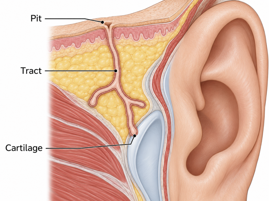
A preauricular sinus is a small congenital opening at the front edge of the upper ear. It is less a dent than a doorway. The pinhole leads into a narrow, skin-lined tunnel under the surface, sometimes short, sometimes long and branching [Children’s Hospital of Philadelphia, Preauricular Pits, 2026]. It forms when the tissue mounds that build the outer ear in the sixth week of pregnancy do not fuse completely [Weinberg, Preauricular Sinuses, 2020].
Because the tunnel is lined with skin, it keeps shedding debris inward. That is where the faint smell or cheesy discharge some people notice comes from. In the Korean survey, 79.08% of cases were on one side only. A pit on its own is not linked to hearing loss. In roughly 3 to 10% of cases, though, it is part of a syndrome such as branchio-oto-renal syndrome, so children who also have other ear, neck, or kidney anomalies may need hearing and kidney checks [Scheinfeld, The preauricular sinus: a review of its clinical presentation, treatment, and associations, 2004].
When should you suspect a preauricular pit infection?
An infected pit swells, hurts, turns red, or leaks pus, sometimes with a fever [Children’s Hospital of Philadelphia, Preauricular Pits, 2026].
The swelling does not always sit on the pit, and that is what makes it easy to miss. Infection can travel along the tunnel and come up on the cheek. In one series from Great Ormond Street Hospital, every infected case involved the cheek skin [Bulstrode, Management of the infected preauricular sinus, 2023].
Why a preauricular sinus infection is so often missed
No study directly counts how many infected pits go unrecognized. What the literature does show is several points where the diagnosis tends to slip.
Common and usually silent
Most preauricular sinuses never cause symptoms [Scheinfeld, The preauricular sinus: a review of its clinical presentation, treatment, and associations, 2004]. In a hospital-based study from Nigeria, 21 of 184 sinuses (11.4%, about one in nine) had progressed to an abscess [Adegbiji, Presentation of preauricular sinus and preauricular sinus abscess in southwest Nigeria, 2013]. Hospital patients are a selected group, so the rate in the general population is probably different. And when a lump finally appears, few people connect it to a pit they have carried without trouble for years.
Mistaken for a boil
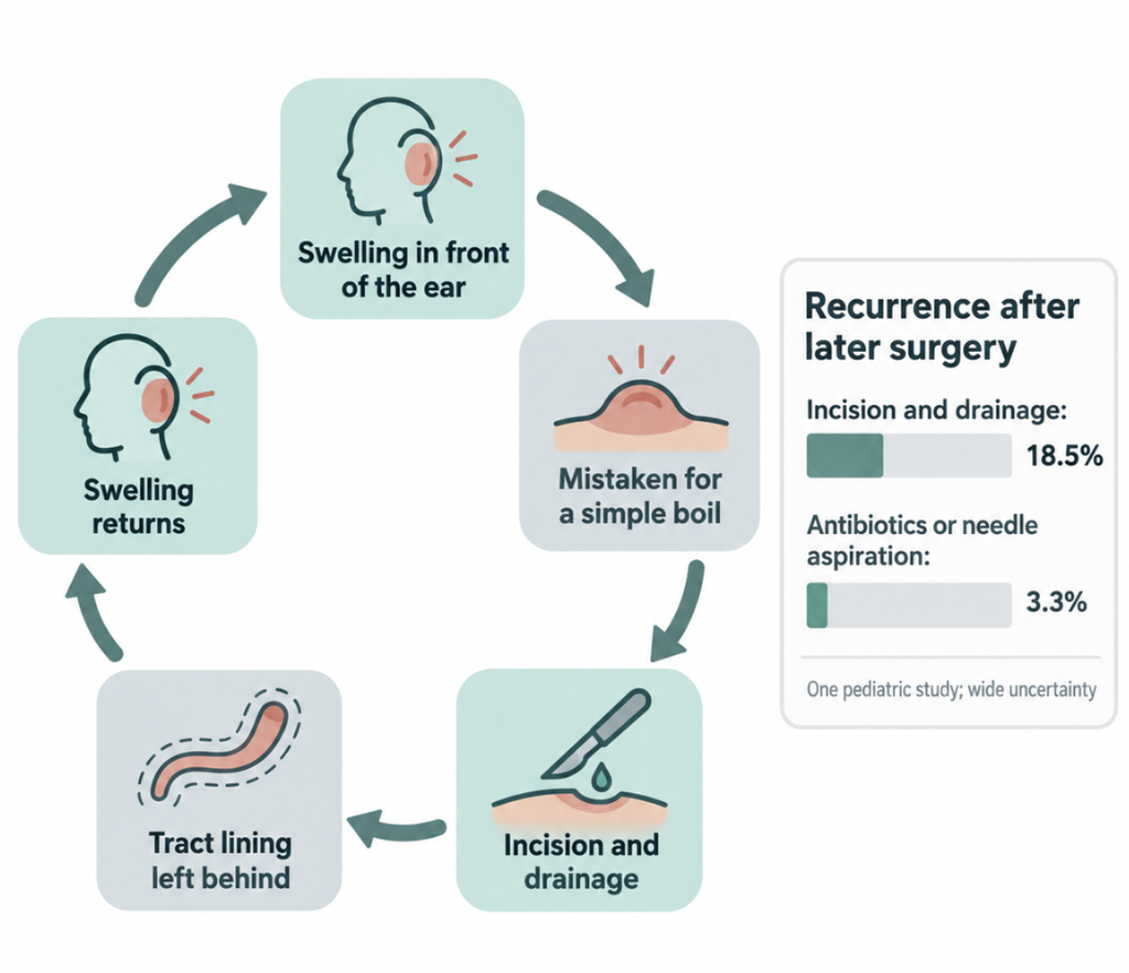
In the same study, referring doctors labeled 71.4% of infected pits as an ordinary ear abscess or cellulitis. Only 9.5% were called a preauricular sinus abscess. The sample was small, so these percentages shouldn’t be over-read. In the Great Ormond Street series, all eight infected children had already been through at least one drainage that didn’t work [Bulstrode, Management of the infected preauricular sinus, 2023].
Why lancing it can backfire
Opening a boil drains a closed pocket, and that is usually the end of it. An infected preauricular sinus is a pocket attached to a tunnel. Cutting into it can scatter the lining that a surgeon later needs to trace. In a study of 69 children, recurrence after the final surgery was 18.5% when the earlier infection had been treated with incision and drainage, compared with 3.3% when it was treated with antibiotics or needle aspiration alone [Rataiczak, Association of recurrence of infected congenital preauricular cysts following incision and drainage vs fine-needle aspiration or antibiotic treatment, 2017]. The uncertainty around that gap was wide, and another series found no link between infection at the time of surgery and recurrence [Gan, Preauricular sinuses in the pediatric population: techniques and recurrence rates, 2013]. So the evidence is a reason for caution. It does not prove that drainage causes harm.
How is it treated, and how well does surgery work?
During an active infection
Oral antibiotics come first. When an abscess forms, many ENT surgeons prefer needle aspiration and keep incision for cases that don’t respond. In a 16-year series from a single surgeon, where infections were managed mainly with antibiotics and sometimes aspiration, only one of 28 infected sinuses recurred after surgery [Isaacson, Comprehensive management of infected preauricular sinuses/cysts, 2019].
The definitive cure: removing the whole tunnel
Antibiotics calm the infection, but the tunnel stays. Complete excision is the only definitive cure. Reported recurrence after excision ranges from 0 to 42% [Bruijnzeel, A systematic review on the surgical outcome of preauricular sinus excision techniques, 2016], and much of that spread comes down to technique.
When tissue from revision operations was examined, 73.7% of specimens still contained skin-lined tract, mostly attached to cartilage, an average of 50.4 months after the first surgery [Kim, Causes and prevention of revision surgery for preauricular sinus: a histopathological analysis, 2019]. Most recurrences, in other words, come from tract left behind the first time, and they can take about four years to show up.
Recurrence by surgical approach
| Approach | What is removed | Pooled recurrence |
|---|---|---|
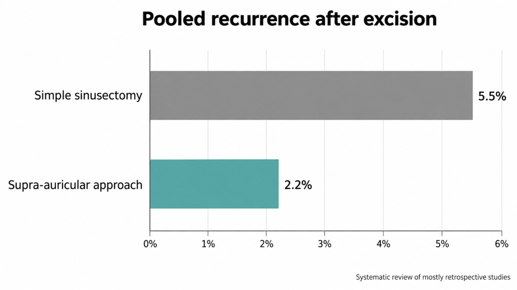
| Simple sinusectomy | Pit and the tract as traced | 5.5% |
| Supra-auricular approach | Wider block down to the temporalis fascia, with adjacent cartilage | 2.2% |
These pooled figures come from a systematic review of mostly retrospective studies, so the gap between them is worth taking seriously but isn’t settled [Bruijnzeel, A systematic review on the surgical outcome of preauricular sinus excision techniques, 2016]. Magnification also matters. Microscope-guided sinusectomy had a recurrence rate of 1.9% in that review, and a 782-ear series linked magnification to fewer recurrences. The same series found no clear benefit from dye staining or cartilage removal alone [Li, Surgical treatment of preauricular fistulas: a 12-year single-center clinical observation, 2023].
If your ear pit has ever been infected, consider removal
A pit that has never been infected can be left alone. After an infection, the picture changes. The tract is still there and still collecting debris, and recurrent infection is a clear indication for complete excision [Scheinfeld, The preauricular sinus: a review of its clinical presentation, treatment, and associations, 2004]. Surgery planned after the inflammation settles is more controlled than an operation forced by a flare.
Practical steps:
- Tell any doctor treating a lump near your ear that you have a pit.
- Don’t squeeze it.
- If an abscess forms, ask whether needle aspiration is an option before incision.
Clinical Perspective. In adults, a swollen lump in front of the ear is easily treated as a sebaceous cyst or a boil, and the pinhole a few millimeters away goes unnoticed. The most useful question is often the simplest one: “Have you always had a small hole here?” If the answer is yes and there has already been an infection, the tract won’t close on its own. Complete removal is what changes the long-term outlook.
Key Takeaways
- A preauricular sinus is a congenital, skin-lined tunnel marked by a pinhole in front of the ear. About 1 in 50 Koreans have one.
- Most pits never cause trouble and need no treatment.
- An infected pit is easily mistaken for a simple boil, and incision and drainage may raise the chance of recurrence after later surgery.
- Complete excision is the only definitive cure. Pooled recurrence is lower after the supra-auricular approach (2.2%) than after simple sinusectomy (5.5%).
- If your pit has ever been infected, talk to an otolaryngologist about planned removal.
FAQ
Why does my preauricular pit keep getting infected?
Because the tract is still there. Its lining keeps shedding debris that bacteria grow in, and antibiotics settle each flare without removing the tract [Scheinfeld, The preauricular sinus: a review of its clinical presentation, treatment, and associations, 2004].
Does a preauricular sinus need surgery?
Not always. A pit that has never been infected can simply be watched. Once infections start, and especially when they keep coming back, removing the entire tract is recommended [Children’s Hospital of Philadelphia, Preauricular Pits, 2026].
Should an infected preauricular pit be drained?
Sometimes, but carefully. Needle aspiration is often preferred, because in one pediatric study incision and drainage was linked to higher recurrence after later surgery [Rataiczak, Association of recurrence of infected congenital preauricular cysts following incision and drainage vs fine-needle aspiration or antibiotic treatment, 2017].
Is a preauricular sinus the same as a perilymphatic fistula?
No. A perilymphatic fistula is a tiny leak between the middle ear and the fluid-filled inner ear. It usually follows head trauma or a pressure injury and causes ear fullness, dizziness, or hearing changes [Vestibular Disorders Association, Perilymph Fistula, 2014]. The names sound alike, but they are different conditions.
References
- Lee KY, Woo SY, Kim SW, Yang JE, Cho YS. The prevalence of preauricular sinus and associated factors in a nationwide population-based survey of South Korea. Otol Neurotol. 2014;35(10):1835-8.
- Children’s Hospital of Philadelphia. Preauricular pits [Internet]. Philadelphia (PA): Children’s Hospital of Philadelphia; 2026 [cited 2026 Sep 25]. Available from: https://www.chop.edu/conditions-diseases/preauricular-pits
- Weinberg J. Preauricular sinuses [Internet]. Medscape; 2020 [cited 2026 Sep 25]. Available from: https://emedicine.medscape.com/article/1118768-overview
- Scheinfeld NS, Silverberg NB, Weinberg JM, Nozad V. The preauricular sinus: a review of its clinical presentation, treatment, and associations. Pediatr Dermatol. 2004;21(3):191-6.
- Bulstrode N, Thacoor A. Management of the infected preauricular sinus. J Plast Reconstr Aesthet Surg. 2023;83:305-7.
- Adegbiji WA, Alabi BS, Olajuyin OA, Nwawolo CC. Presentation of preauricular sinus and preauricular sinus abscess in southwest Nigeria. Int J Biomed Sci. 2013;9(4):260-3.
- Rataiczak H, Lavin J, Levy M, Bedwell J, Preciado D, Reilly BK. Association of recurrence of infected congenital preauricular cysts following incision and drainage vs fine-needle aspiration or antibiotic treatment: a retrospective review of treatment options. JAMA Otolaryngol Head Neck Surg. 2017;143(2):131-4.
- Gan EC, Anicete R, Tan HK, Balakrishnan A. Preauricular sinuses in the pediatric population: techniques and recurrence rates. Int J Pediatr Otorhinolaryngol. 2013;77(3):372-8.
- Isaacson G. Comprehensive management of infected preauricular sinuses/cysts. Int J Pediatr Otorhinolaryngol. 2019;127:109682.
- Bruijnzeel H, van den Aardweg MT, Grolman W, Stegeman I, van der Veen EL. A systematic review on the surgical outcome of preauricular sinus excision techniques. Laryngoscope. 2016;126(7):1535-44.
- Kim WJ, Lee YM, Kim DH, Choe S, Lee D, Park SY, et al. Causes and prevention of revision surgery for preauricular sinus: a histopathological analysis. Int J Pediatr Otorhinolaryngol. 2019;116:199-203.
- Li K, Hao Y, Zhao J, Zhou L, Wu Y, Zeng X, et al. Surgical treatment of preauricular fistulas: a 12-year single-center clinical observation. BMC Surg. 2023;23(1):297.
- Vestibular Disorders Association. Perilymph fistula [Internet]. Portland (OR): VeDA; 2014 [cited 2026 Sep 25]. Available from: https://royalwolverhampton.nhs.uk/wp-content/uploads/2025/06/Perilymph-Fistula.pdf
Joonpyo Hong, MD is a board-certified otolaryngologist practicing in Korea. This article reflects his clinical interpretation of published research and does not constitute individual medical advice.
For more articles:
https://curiousmd.com/perilymphatic-fistula-from-weightlifting/
https://curiousmd.com/carotid-blowout-syndrome-greys-anatomy/
https://curiousmd.com/facial-and-neck-lacerations-by-site/
Link out to:
https://www.chop.edu/conditions-diseases/preauricular-pits — Children’s Hospital of Philadelphia’s patient guide to preauricular pits, infection, and when complete removal is recommended.
https://emedicine.medscape.com/article/1118768-overview — Medscape clinical reference on the anatomy, epidemiology, and complications of preauricular sinuses.
https://pedemmorsels.com/preauricular-pits-and-infection/ — An emergency physician’s summary of why infected pits should not be treated like simple abscesses.
https://royalwolverhampton.nhs.uk/wp-content/uploads/2025/06/Perilymph-Fistula.pdf — Vestibular Disorders Association explainer on perilymph fistula, a different inner-ear condition with a similar name.