
Picture a 20-year-old on day 3 of fever and sore throat; the first question is infectious mononucleosis vs tonsillitis. The tonsils are coated, both sides of the neck are swollen, and the blood test that could confirm mono will not be back for days. This article covers how to tell the two apart, why early testing often misleads, and what you can start while results are pending.
Infectious mononucleosis vs tonsillitis: clues in the history and exam
The mouth alone is a poor guide. Tonsillar exudate appears in both conditions, including about half of mono cases, so it tells you little [Sylvester, Infectious Mononucleosis: Rapid Evidence Review, 2023].
Age is the first clue. Mono peaks at ages 15 to 24, and about 1 in 13 sore-throat patients aged 16 to 20 turns out to have it [Ebell, Does This Patient Have Infectious Mononucleosis? The Rational Clinical Examination Systematic Review, 2016].
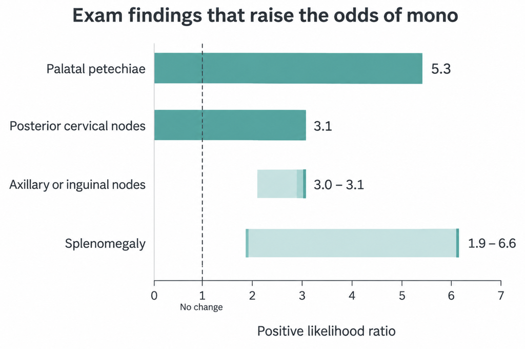
The neck and body exam carry more weight than the throat:
| Finding | Positive likelihood ratio |
|---|---|
| Palatal petechiae | 5.3 |
| Posterior cervical nodes | 3.1 |
| Axillary or inguinal nodes | 3.0 to 3.1 |
| Splenomegaly | 1.9 to 6.6 |
Posterior cervical nodes roughly triple the odds. Absence counts too: lymphadenopathy is present in 91% of cases, so a patient with no enlarged nodes anywhere is less likely to have mono. A later meta-analysis agreed and found blood counts more accurate than symptoms and signs [Cai, Accuracy of Signs, Symptoms, and Hematologic Parameters for the Diagnosis of Infectious Mononucleosis: A Systematic Review and Meta-Analysis, 2021].
A blood count with differential is the cheapest useful test. Atypical lymphocytes of 10% or more carry a likelihood ratio of 11.4, higher than most physical signs [Ebell, Does This Patient Have Infectious Mononucleosis? The Rational Clinical Examination Systematic Review, 2016].
ENT clinics have one more clue. In one small study, endoscopy showed swollen nasopharyngeal lymphoid tissue covered with fibrinous membranes in 24 of 26 IM patients and in 0 of 24 with tonsillitis [Weber, Nasopharyngeal endoscopy adds to reliability of clinical diagnosis of infectious mononucleosis, 2001]. Larger studies are needed, but if you already perform endoscopy, the extra look costs nothing.
The mono test problem: accurate, but not on day 3
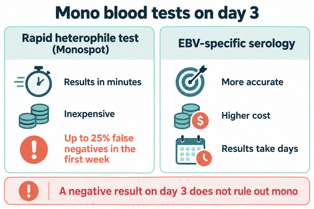
The rapid heterophile antibody test (Monospot) is fast and inexpensive. Overall it has a sensitivity of 87% and specificity of 91%, but it gives up to 25% false negatives in adults in the first week and can miss mono in children younger than five [Sylvester, Infectious Mononucleosis: Rapid Evidence Review, 2023]. On day 3, a negative result does not rule out mono.
EBV-specific antibody testing (viral capsid antigen IgM and IgG, with nuclear antigen antibody) performs better, with sensitivity 97% and specificity 94%. It costs more, and results often take days rather than minutes. Most general outpatient clinics cannot run it in-house, so the sample goes to an outside lab and the patient waits.
Because treatment is supportive, a confirmed result rarely changes that day’s prescription. It mainly changes what you tell the patient about the spleen and the expected course.
Waiting for results: what you can treat on day 3
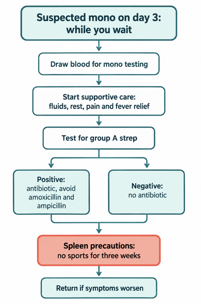
The decision that cannot wait is whether a bacterial infection is present, and that calls for a group A streptococcal test. Antibiotics are reasonable only when streptococcal infection is confirmed alongside mono.
Supportive care can start the same day: fluids, rest, and an analgesic for fever and throat pain. Use acetaminophen judiciously because mono often inflames the liver, and avoid aspirin in children and adolescents [Sylvester, Infectious Mononucleosis: Rapid Evidence Review, 2023].
Older reports put the rash rate after ampicillin at 80% to 100%. A study of 238 children found amoxicillin-associated rash in 29.5%, and rash rates of 32.9% treated vs 23.1% untreated were not significantly different [Chovel-Sella, Incidence of rash after amoxicillin treatment in children with infectious mononucleosis, 2013]. A larger cohort of 767 children across 14 hospitals found antibiotic-related rash in 4.08% with amoxicillin vs 8.15% with other antibiotics [Zhang, Association between Antibiotic Exposure and the Risk of Rash in Children with Infectious Mononucleosis: a Multicenter, Retrospective Cohort Study, 2023]. The two studies used different populations and methods, so their figures are not directly comparable. Both suggest a smaller amoxicillin effect than textbooks, and even some current reviews, describe. Still, avoiding aminopenicillins when mono is suspected costs little.
A Cochrane review of 7 trials and 362 participants found sore throat relief at 12 hours in two trials, which did not last, and no benefit in 8 of 10 assessments [Rezk, Steroids for symptom control in infectious mononucleosis, 2015]. Steroids are reserved for threatened airway obstruction or severe disease, and antivirals are not recommended.
One precaution should start before confirmation. Splenic rupture is rare, at 0.1% to 0.2%, but 73.8% of cases occur within 21 days of onset. Current guidance advises no athletic activity for three weeks from symptom onset [Sylvester, Infectious Mononucleosis: Rapid Evidence Review, 2023].
Can neck ultrasound point to mono?
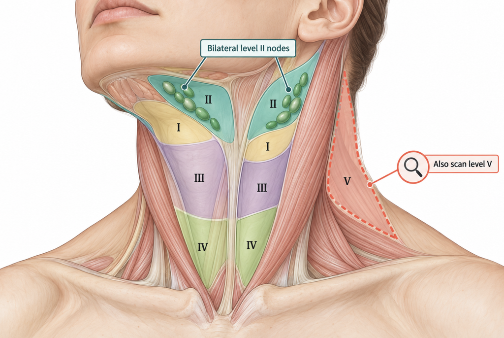
In a comparison study, nodes from mono patients were bilateral in 91%, matted in 83%, matted on both sides in 66%, had indistinct margins in 80%, were round in 69% and showed central hilar flow in 89.8% [Fu, Sonographic appearance of cervical lymphadenopathy due to infectious mononucleosis in children and young adults, 2014]. Bilateral matting was the most specific sign, though many patients lacked it. Indistinct margins were more common in mono than in bacterial lymphadenitis.
In 61 children with mono, enlargement was symmetric and all enlarged nodes sat in level II, the upper jugular chain [Bu, Ultrasound characteristics of cervical lymph nodes in children with infectious mononucleosis, 2025]. The study had no comparison group, so it describes how mono looks but cannot show that these findings separate mono from other causes.
Clinical Perspective. Posterior cervical swelling is the best-known sign of mono, so sweep the posterior triangle (level V) deliberately instead of stopping at the upper jugular chain. Bilateral enlarged level V nodes on ultrasound correspond to the palpation sign, which roughly triples the odds on examination, and ultrasound can find nodes too small to feel. In the pediatric study above, enlargement was confined to level II, so this targeted view needs testing, especially in adolescents and adults.
Kawasaki disease produces clustered, grape-like nodes that resemble acute EBV infection [Tashiro, Ultrasonographic evaluation of cervical lymph nodes in Kawasaki disease, 2002], and tuberculous nodes can also mat. Ultrasound findings can raise or lower suspicion of mono but cannot confirm the diagnosis.
Key takeaways
- Posterior cervical nodes, palatal petechiae and splenomegaly raise the likelihood of mono; tonsillar exudate does not help.
- The heterophile test misses up to 25% of adult cases in the first week, so an early negative does not exclude mono.
- Mono treatment is supportive; the urgent decision is whether group A strep needs antibiotics.
- Recent studies suggest amoxicillin rash is far rarer than the historical 80% to 100%, but avoiding aminopenicillins remains sensible.
- Bilateral, matted level II nodes on ultrasound point toward mono but overlap with other causes.
FAQ
How do you tell mono from tonsillitis?
Check the neck. Posterior cervical, axillary or inguinal nodes, palatal petechiae, an enlarged spleen and atypical lymphocytes on a blood count all make mono more likely, while exudate on the tonsils appears in both [Ebell, Does This Patient Have Infectious Mononucleosis? The Rational Clinical Examination Systematic Review, 2016].
Is the monospot test accurate in the first week?
No. It misses up to 25% of adult cases in the first week, so an early negative may need repeating or EBV-specific testing [Sylvester, Infectious Mononucleosis: Rapid Evidence Review, 2023].
What can you take for mono while waiting for test results?
Supportive care. Fluids, rest and an analgesic for fever and pain are the treatment; routine steroids and antivirals are not recommended [Rezk, Steroids for symptom control in infectious mononucleosis, 2015].
Should I avoid amoxicillin if mono is suspected?
Usually, yes. Rash after amoxicillin is less common than once believed, but antibiotics are needed only if a bacterial infection such as strep is confirmed [Chovel-Sella, Incidence of rash after amoxicillin treatment in children with infectious mononucleosis, 2013].
Can a neck ultrasound show mono?
Partly. Bilateral, matted nodes fit mono, but the findings overlap with other conditions [Fu, Sonographic appearance of cervical lymphadenopathy due to infectious mononucleosis in children and young adults, 2014].
References
- Ebell MH, Call M, Shinholser J, Gardner J. Does This Patient Have Infectious Mononucleosis?: The Rational Clinical Examination Systematic Review. JAMA. 2016;315(14):1502-9.
- Cai X, Ebell MH, Haines L. Accuracy of Signs, Symptoms, and Hematologic Parameters for the Diagnosis of Infectious Mononucleosis: A Systematic Review and Meta-Analysis. J Am Board Fam Med. 2021;34(6):1141-1156.
- Sylvester JE, Buchanan BK, Silva TW. Infectious Mononucleosis: Rapid Evidence Review. Am Fam Physician. 2023;107(1):71-78.
- Weber R, Hegenbarth V, Kaftan H, Krüpe H, Jaspersen D, Keerl R. Nasopharyngeal endoscopy adds to reliability of clinical diagnosis of infectious mononucleosis. J Laryngol Otol. 2001;115(10):792-5.
- Chovel-Sella A, Ben Tov A, Lahav E, Mor O, Rudich H, Paret G, et al. Incidence of rash after amoxicillin treatment in children with infectious mononucleosis. Pediatrics. 2013;131(5):e1424-7.
- Zhang R, Mao Z, Xu C, Wang W, Kwong JS, Xu M, et al. Association between Antibiotic Exposure and the Risk of Rash in Children with Infectious Mononucleosis: a Multicenter, Retrospective Cohort Study. Antimicrob Agents Chemother. 2023;67(6):e0024923.
- Rezk E, Nofal YH, Hamzeh A, Aboujaib MF, AlKheder MA, Al Hammad MF. Steroids for symptom control in infectious mononucleosis. Cochrane Database Syst Rev. 2015;2015(11):CD004402.
- Fu XS, Guo LM, Lv K, Wang L, Ran WQ, Tan QT, et al. Sonographic appearance of cervical lymphadenopathy due to infectious mononucleosis in children and young adults. Clin Radiol. 2014;69(3):239-45.
- Bu X, Sun S, Zhang Y, Yuan T, Wang C. Ultrasound characteristics of cervical lymph nodes in children with infectious mononucleosis. Med Ultrason. 2025;28(1):38-42.
- Tashiro N, Matsubara T, Uchida M, Katayama K, Ichiyama T, Furukawa S. Ultrasonographic evaluation of cervical lymph nodes in Kawasaki disease. Pediatrics. 2002;109(5):E77.
Joonpyo Hong, MD is a board-certified otolaryngologist practicing in Korea. This article reflects his clinical interpretation of published research and does not constitute individual medical advice.
For more articles:
https://curiousmd.com/sore-throat-on-one-side-peritonsillar-abscess/
https://curiousmd.com/tonsil-stones-do-you-need-surgery/
https://curiousmd.com/aphthous-ulcer-treatment-ent-guide/
Link out to:
https://www.aafp.org/afp/2023/0100/infectious-mononucleosis — American Academy of Family Physicians evidence review covering diagnosis, testing and supportive treatment of infectious mononucleosis.
https://www.cochrane.org/evidence/CD004402_steroids-short-term-symptom-control-infectious-mononucleosis-glandular-fever — Cochrane plain-language summary on whether steroids help control symptoms of glandular fever.
https://medlineplus.gov/ency/article/000591.htm — U.S. National Library of Medicine patient guide to mono, including rash with amoxicillin and spleen precautions.
https://nidirect.gov.uk/conditions/glandular-fever — UK public health guide to glandular fever symptoms, blood tests and self-care.