
Three patients walk into a voice clinic, all hoarse, all with a small bump on a vocal fold — and the right call in a vocal nodule vs polyp vs cyst decides everything that follows. One will recover with voice therapy alone. One might, if it’s the right kind of polyp. The third — the one with a cyst — almost certainly needs surgery, and no amount of voice rest will change that.
Those three outcomes come from three lesions that look deceptively similar to the untrained eye: the nodule, the polyp, and the intracordal cyst (which itself splits into mucous and epidermoid types). Telling them apart is not academic hair-splitting. It changes what treatment works, how long recovery takes, and whether a patient should expect surgery at all. Here is how they differ, and what the evidence says about treating each one.
The four lesions at a glance
| Nodule | Polyp | Mucous retention cyst | Epidermoid cyst | |
|---|---|---|---|---|
| Main cause | Chronic vocal overuse | Overuse, or a single vocal injury | Blocked mucous-gland duct | Congenital cell rest or buried epithelium after microtrauma |
| Overuse link | Strong | Strong | Weak / often unrelated | Variable |
| Sides affected | Bilateral (both folds) | Usually one fold | One fold | One fold |
| Contents | Callus-like fibrous tissue | Fluid, vascular, or gelatinous | Trapped mucus (clear–yellow) | Keratin (pearly white) |
| Mucosal wave | Reduced | Often preserved | Absent | Absent |
| Resolves with voice rest? | Often | Sometimes | Rarely | No |
| First-line treatment | Voice therapy | Voice therapy trial, then surgery | Surgery | Surgery |
Nodules: the bilateral “calluses”
Vocal fold nodules form where the folds collide hardest — the midpoint of the membranous cord — during repeated, forceful voice use. Because both folds strike each other in the same place, nodules almost always appear on both sides, facing each other like mirror images. They are essentially calluses, and like calluses on a hand, early soft nodules can shrink or disappear once the friction stops.
This is why teachers, singers, coaches, and cheerleaders sit at the top of the risk list. The tissue itself is fibrous and surface-level, which matters enormously for treatment: a surface reaction to behavior tends to respond to changing the behavior.
Polyps: one-sided, sometimes sudden
A polyp usually forms on one fold and can take many shapes — a broad-based bump, a stalked lesion, or a blood-filled blister. Unlike nodules, which need chronic overuse, a polyp can appear after a single traumatic event, such as screaming at a concert or a violent coughing fit. Some polyps are vascular and gelatinous; others become fibrotic and firm over time.
That variability predicts treatment response. A soft, translucent polyp behaves very differently from a hardened, hemorrhagic one — a distinction that becomes important below.
Intracordal cysts: mucous vs epidermoid
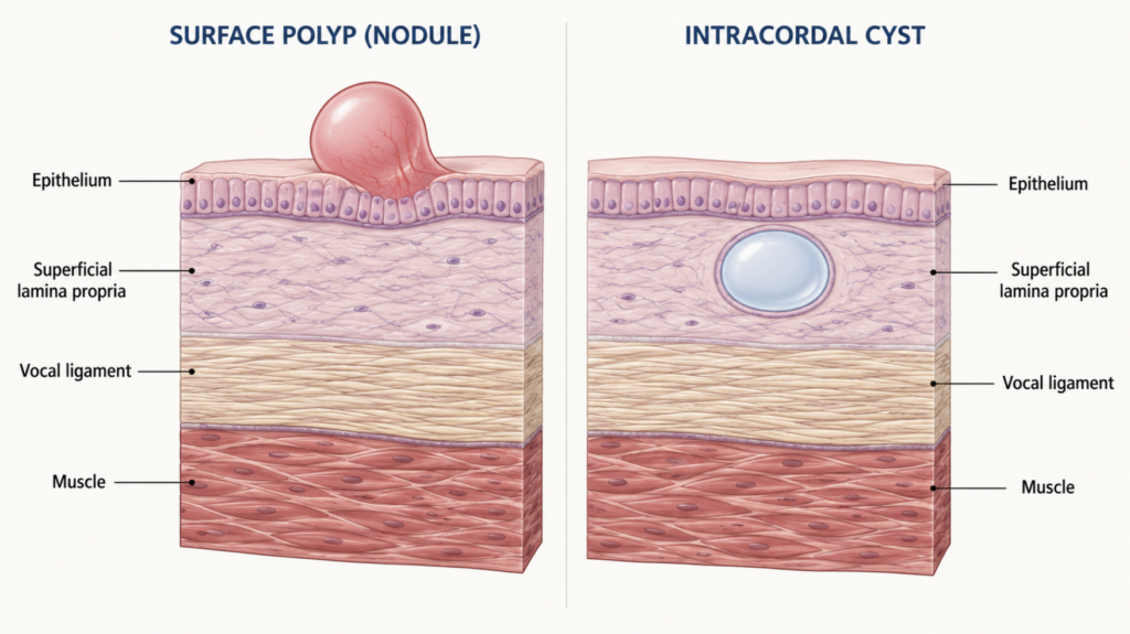
This is where most general explanations stop short. A cyst is not a surface swelling but a contained sac buried within the superficial layer of the vocal fold (the lamina propria). Because it sits inside the fold rather than on top of it, resting the voice does not drain it. There are two kinds, and they arise for entirely different reasons.
A mucous retention cyst forms when one of the small mucous glands just beneath the fold’s edge becomes plugged. Mucus keeps being produced but can no longer drain, so it accumulates into a fluid-filled sac that is typically clear or yellowish. These often have little to do with vocal overuse.
An epidermoid (keratin) cyst is filled with keratin rather than mucus, giving it a classically pearly white appearance. These are thought to arise either from congenital nests of epithelial cells left behind during development, or from surface epithelium that gets buried in the fold after microtrauma. They tend to be smaller, subtler, and more stubborn.
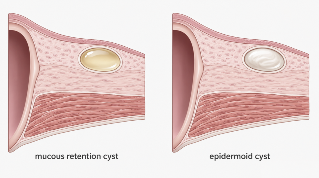
One caveat on appearance: the familiar teaching that mucous cysts look translucent and epidermoid cysts look opaque is only a rough guide. A clinicopathologic study of 69 excised cysts found that a cyst’s opacity did not reliably predict its subtype, though epidermoid cysts were more likely to show clinical inflammation — meaning the definitive answer comes from histology after removal, not from the exam-room view.
Both types share one crucial feature: because they distort the fold from the inside, they flatten or abolish the mucosal wave — the rippling motion of the fold’s cover during vibration. That single finding is often what separates a cyst from a polyp at the bedside.
Vocal Nodule vs Polyp vs Cyst: What Sets Them Apart
All four are benign, all cause hoarseness, and all can trace back — at least partly — to how the vocal folds meet during voice use. The differences that matter clinically are three: how many folds are involved (nodules bilateral; polyps and cysts usually unilateral), where the lesion sits (nodules and polyps mainly on the surface and partly the lamina propria; cysts inside the lamina propria), and what happens to the mucosal wave (preserved in polyps, absent in cysts). On a mirror exam these lesions can masquerade as one another, which is why videostroboscopy — a strobe-light exam that visualizes fold vibration — is the key differentiator, and why a cyst’s exact subtype is often confirmed only during surgery.
Treatment: what actually works, lesion by lesion
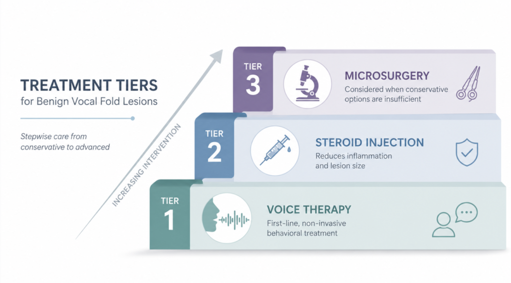
Three tiers of treatment apply to all four lesions, but the balance shifts by type. A useful rule of thumb: the more a lesion is a surface reaction to overuse (nodule, then polyp), the more behavioral therapy alone can resolve it; the more it is a contained structural mass inside the fold (cyst), the more surgery is required.
Behavioral treatment: voice therapy and voice rest
Voice therapy — delivered by a speech-language pathologist — combines vocal hygiene, relative voice rest, elimination of phonotrauma (yelling, throat-clearing), and direct retraining of how the voice is produced.
For nodules, it is first-line and frequently curative. A 2024 systematic review concluded that voice therapy is the preferred non-invasive treatment, though it takes longer to work and, in children, is limited by compliance; notably, hard or mature nodules responded better to surgery than to therapy.
For polyps, a therapy trial is worthwhile but succeeds only about half the time. In a retrospective series of 57 patients with polyps and cysts, 49.1% achieved symptom resolution with voice therapy alone — but the type of polyp mattered enormously: translucent polyps responded 81.8% of the time, versus only 15.4% for fibrotic and 25.0% for hemorrhagic polyps. A 2023 meta-analysis found voice therapy, surgery, and combined treatment were all highly effective for polyps, with phonosurgery and combined treatment providing the greatest overall improvement.
For cysts, therapy alone rarely resolves the lesion, because you cannot rehabilitate away a fluid- or keratin-filled sac. Its real role is peri-operative: preparing the voice before surgery and rebuilding healthy technique afterward to prevent recurrence.
Clinical perspective: The single most useful bedside clue is the mucosal wave on videostroboscopy. If it’s preserved, think polyp and a therapy trial is reasonable. If it’s absent over a unilateral mass, think cyst — and counsel the patient early that voice rest won’t be enough.
Pharmacologic treatment
The “injection” that actually treats these lesions is intralesional (vocal-fold) steroid injection — an office-based procedure, done under local anesthesia, distinct from injection laryngoplasty discussed below.
A randomized trial in small (≤5 mm) benign lesions found that intralesional steroid injection significantly reduced lesion size at 12 weeks with no recurrence, offering an office alternative to microsurgery for small lesions. For polyps specifically, a percutaneous corticosteroid injection series of 24 patients completed the procedure in 22, typically within 20 minutes, with a 91% response rate and 59% complete remission — all while avoiding general anesthesia and the scarring risk of open surgery.
The honest caveat is durability. A 2-year prospective surveillance of 189 patients found that among those who initially responded, steroid injection remained effective at two years in 54% of polyps, 49% of nodules, and only 43% of mucus-retention cysts — well below what surgery achieves. In other words, injection can buy time and sometimes avoid surgery, but relapse within two years is common, especially for cysts.
A second class of “medication” doesn’t target the lesion at all — it targets the drivers. Chronic cough and throat-clearing inflict repeated shear injury, so cough-suppression strategies and, where relevant, antitussives are used as adjuncts. Reflux is a documented contributor: a meta-analysis found that objectively diagnosed laryngopharyngeal reflux was associated with roughly three-fold higher odds of benign vocal fold lesions, which is why reflux control is a standard part of management. These medications treat causes, not the bump itself.
Surgical treatment
Laryngeal microsurgery (LMS) — also called phonomicrosurgery or microlaryngoscopy — is the definitive option for lesions that fail conservative care and for essentially all cysts. The guiding principle is preservation: removing the lesion while sparing the maximum amount of the superficial lamina propria, because that layer is what lets the fold vibrate. Sacrifice it and you trade a lesion for a stiff, scarred fold.
For cysts, surgery is primary. The goal is to remove the intact sac; if part of the cyst wall is left behind, mucous cysts in particular can re-accumulate and require revision surgery. For polyps, surgery is the standard when a therapy trial fails or when the lesion is large, vascular, or fibrotic. For nodules, surgery is the exception, reserved for firm, mature nodules that don’t respond to therapy.
Surgery and injection aren’t mutually exclusive. In a cohort of 211 patients, adding an immediate intralesional steroid injection at the end of microsurgery lowered the rate of recurrent lesions and reduced the risk of persistent hoarseness afterward.
Two more surgical tools deserve a clarifying word. Angiolytic (KTP) laser is well suited to vascular polyps and can often be done in the office; it targets the lesion’s blood supply while sparing surrounding mucosa. Injection laryngoplasty, on the other hand, is frequently confused with intralesional steroid injection but does something completely different: it augments a fold with a filler (hyaluronic acid, collagen, or fat) to close a glottic gap in conditions like vocal fold paralysis. It is not a treatment for nodules, polyps, or cysts themselves — it addresses vocal-fold closure, not the lesion. Conflating the two is one of the more common misunderstandings patients bring to the clinic.
Treatment-by-lesion quick reference
| Lesion | Behavioral therapy | Steroid injection | Microsurgery |
|---|---|---|---|
| Nodule | First-line, often curative | Effective; ~49% at 2 yrs (initial responders) | Only if firm/refractory |
| Polyp | Worth a trial (~49% resolve; translucent best) | 91% response, 59% remission short-term | Standard if therapy fails |
| Mucous cyst | Peri-operative support only | Weak durability (~43% at 2 yrs) | Primary |
| Epidermoid cyst | Peri-operative support only | Not yet studied* | Primary |
Symptoms and when to see a doctor
All four lesions announce themselves the same way: a hoarse, rough, or breathy voice that often worsens with use and improves with rest — until, with cysts, rest stops helping. Some people notice a voice that cracks, tires by evening, or loses its higher notes. Because the folds no longer close cleanly, extra effort creeps in, and that strain can spiral into muscle-tension patterns that make things worse.
The practical rule: any hoarseness lasting more than two to three weeks without a clear explanation deserves a laryngeal exam. Persistent voice change is also, rarely, how laryngeal cancer presents, and a benign-looking lesion doesn’t rule that out until it resolves or is examined properly. That concern rises with known risk factors — older age, a history of smoking, heavy alcohol use, and unexplained weight loss — which should lower the threshold for prompt evaluation.
Key takeaways
- Nodules are bilateral and overuse-driven; polyps and cysts are usually one-sided.
- Cysts sit inside the vocal fold and rarely resolve without surgery — mucous cysts hold mucus, epidermoid cysts hold keratin.
- Voice therapy is curative-intent for nodules, a fair trial for polyps (best for translucent ones), and supportive-only for cysts.
- Steroid injection gives strong short-term results for polyps and nodules but weaker two-year durability, especially for cysts.
- Injection laryngoplasty treats vocal-fold closure, not the lesion — it is not the same as intralesional steroid injection.
FAQ
Can a vocal cyst go away on its own?
Rarely. Unlike nodules, a cyst is a contained sac buried inside the vocal fold, so voice rest can’t drain it. Most cysts that affect the voice need surgical removal, though voice therapy plays a supporting role before and after.
How is a polyp different from a nodule?
Nodules are bilateral calluses caused by chronic overuse and usually respond to voice therapy. Polyps are typically one-sided, can appear after a single vocal injury, and are more variable — some resolve with therapy, others need surgery.
Does a steroid injection work instead of surgery?
For nodules and small or translucent polyps, it often can, with high short-term response rates and no general anesthesia. The trade-off is durability: relapse within two years is common, particularly for cysts, so surgery remains preferred when a lasting result matters.
What’s the difference between a mucous and an epidermoid cyst?
A mucous retention cyst forms from a blocked gland and holds trapped mucus, appearing clear or yellowish. An epidermoid cyst holds keratin, looks pearly white, is often congenital, and tends to be more stubborn.
References
- Cohen SM, Garrett CG. Utility of voice therapy in the management of vocal fold polyps and cysts. Otolaryngol Head Neck Surg. 2007;136(5):742-746.
- Barsties V Latoszek B, Watts CR, Hetjens S, Neumann K. The efficacy of different voice treatments for vocal fold polyps: a systematic review and meta-analysis. J Clin Med. 2023;12(10):3451.
- Baali MH, Shaheen MH, Khan MF, Neazy AA, Basyuni MA, Altowairqi A. Optimizing management strategies for vocal cord nodules: a systematic review. Cureus. 2024;16(12):e75916.
- Ramavat AS, Tiwana H, Banumathy N, Bakshi J, Panda N, Goel A. Efficacy of intralesional steroid injection in small benign vocal fold lesions. J Voice. 2018;33(5):767-772.
- Hsu YB, Lan MC, Chang SY. Percutaneous corticosteroid injection for vocal fold polyp. Arch Otolaryngol Head Neck Surg. 2009;135(8):776-780.
- Wang CT, Lai MS, Cheng PW. Long-term surveillance following intralesional steroid injection for benign vocal fold lesions. JAMA Otolaryngol Head Neck Surg. 2017;143(6):589-594.
- Cho JH, Kim SY, Joo YH, Park YH, Hwang WS, Sun DI. Efficacy and safety of adjunctive steroid injection after microsurgical removal of benign vocal fold lesions. J Voice. 2017;31(5):615-620.
- Ren QW, Lei G, Zhao YL, Zhou L, Luo XL, Peng SL. Laryngopharyngeal reflux and benign vocal fold lesions: a systematic review and meta-analysis. Otolaryngol Head Neck Surg. 2024;170(2):309-319.
- Alfonso-Ying DA, Clark CM, Scognamiglio T, Rives H, Sulica L. Correlation between laryngoscopic appearance and histopathology in vocal fold cysts. J Voice. 2026;40(1):31-37.
Joonpyo Hong, MD is a board-certified otolaryngologist practicing in Korea. This article reflects his clinical interpretation of published research and does not constitute individual medical advice.
For more interesting content:
https://curiousmd.com/jon-bon-jovi-vocal-cord-surgery-medialization-thyroplasty/
https://curiousmd.com/mdvp-voice-analysis-interpretation/
https://curiousmd.com/ai-laryngeal-cancer-detection/
Link out to
- ASHA — Vocal Cord Nodules and Polyps (American Speech-Language-Hearing Association)
- Penn Medicine — Vocal Cord Lesions: Nodules, Polyps and Cysts
- The Voice Foundation — Understanding Nodules, Polyps, Cysts