
In a study of 1,975 thyroid fine needle aspirations performed in a single Canadian city, the rate of non-diagnostic samples was 3.4% when a pathologist held the needle and 37.9% when a low-volume surgical practice did [Houdek D, Factors Affecting Thyroid Nodule Fine Needle Aspiration Non-Diagnostic Rates: A Retrospective Association Study of 1975 Thyroid Biopsies, 2021]. Same test, same anatomy, an eleven-fold difference in the chance of walking out with no answer at all. Hold onto that gap, because the fine needle aspiration vs core needle biopsy question turns out to matter less than most people expect — and the question of who is holding the needle turns out to matter more.
This article covers what fine needle aspiration biopsy (FNAB) and core needle biopsy (CNB) each sample, why both still exist, whether sampling a fraction of a lesion is a defensible way to diagnose disease, when neither is the right first move, and what the published complication rates actually are.
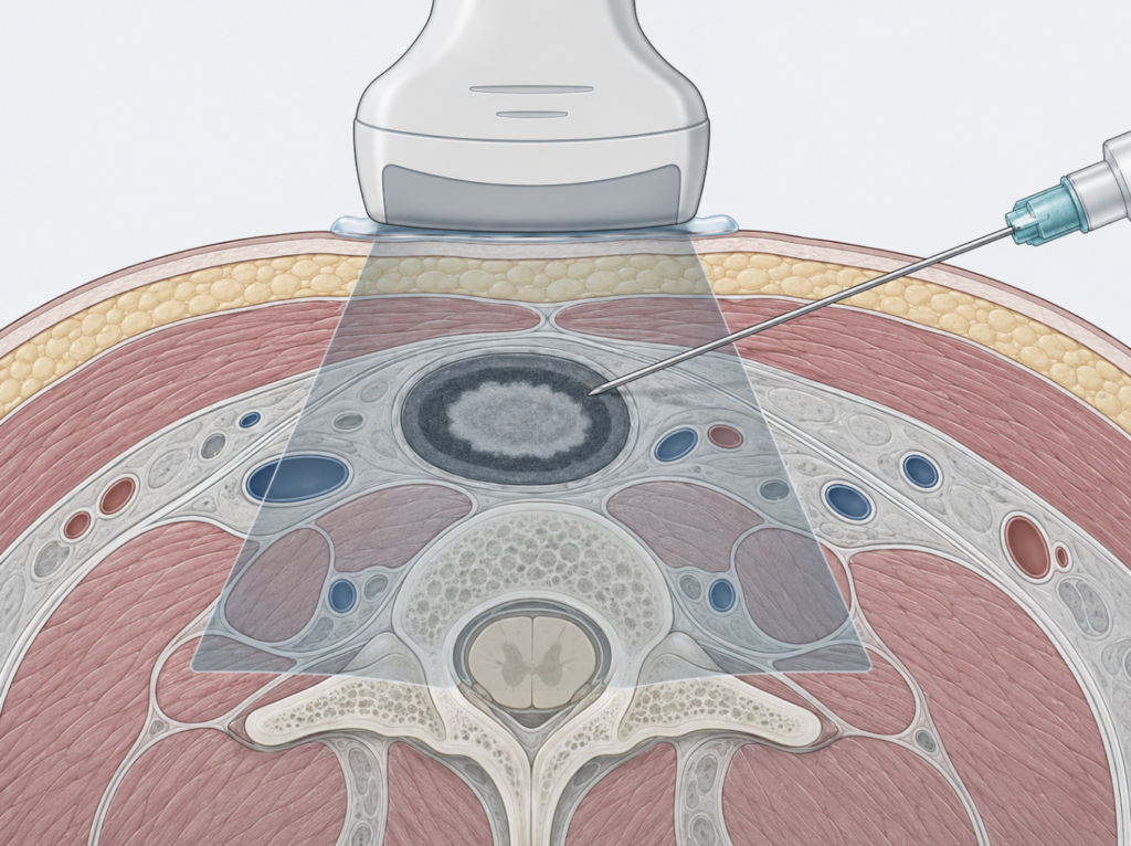
What Fine Needle Aspiration Biopsy and Core Needle Biopsy Actually Are
Fine needle aspiration biopsy: a cell sample
FNAB uses a needle finer than the one used to draw blood — typically 23 to 27 gauge. There is no incision and usually no local anesthetic. The needle is advanced into the lesion and moved back and forth while capillary action, sometimes assisted by suction, pulls individual cells and small cell clusters into the needle hub. Two to four passes are standard.
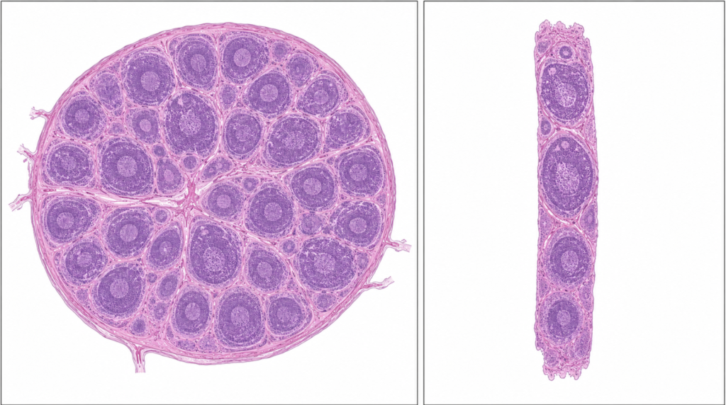
What comes out is cytology: dispersed cells, without the surrounding architecture. A cytopathologist evaluates nuclear size and contour, chromatin pattern, how the cells group themselves, and what sits in the background — colloid, mucin, lymphoid tangles, necrotic debris.
The result is not reported as a yes or no. It is reported inside a tiered framework: the Bethesda System for thyroid, the Milan System for salivary glands. Each tier carries a published probability of malignancy attached to it.
Core needle biopsy: a tissue sample
CNB uses a 16- to 20-gauge spring-loaded cutting needle. Local anesthetic is required, and a small nick is made in the skin. Each firing retrieves an intact cylinder of tissue roughly 1 to 1.5 cm long.
What comes out is histology. The tissue architecture survives — how cells sit relative to one another, whether a capsule exists, whether anything has breached it, what the stroma looks like. That intact block can then be sectioned repeatedly for immunohistochemistry, fluorescence in situ hybridization, and molecular panels.
The distinction worth carrying through the rest of this article is not needle diameter. It is this: FNAB samples cells; CNB samples tissue with its architecture intact.
FNA vs Core Needle Biopsy: Why Two Techniques Instead of One
Some diagnoses are written in the cells. Others are written in the arrangement of the cells.
Papillary thyroid carcinoma is essentially a nuclear diagnosis — the grooves, the pseudoinclusions, the powdery chromatin. Cytology sees it well. Follicular carcinoma is defined by capsular and vascular invasion, which is a statement about geography, not about individual cells. No aspirate, however cellular, can resolve it. That is not an operator failure; it is a structural boundary of the technique.
Lymphoma sits at the far end of that spectrum, requiring nodal architecture plus a battery of ancillary tests before a subtype can be named.
Where FNAB holds its ground
- Thyroid nodules. The entire management algorithm is built on cytology and its risk tiers.
- Cystic lesions, where aspiration is simultaneously diagnostic and therapeutic.
- Confirming metastatic squamous cell carcinoma in a node when a primary is known or suspected. A positive is a positive, and a fine needle is enough to get one.
- Lesions near the facial nerve or great vessels, where a cutting needle is the poorer risk.
- Anticoagulated patients.
- Speed and cost, and — when rapid on-site evaluation is available — an adequacy answer before the patient leaves the room.
Where CNB pulls ahead
A meta-analysis of ultrasound-guided salivary gland CNB across 1,315 procedures found pooled sensitivity of 0.94 and specificity of 0.98, with an area under the summary ROC curve of 0.98 [Kim HJ, Ultrasound-Guided Core Needle Biopsy in Salivary Glands: A Meta-Analysis, 2018]. A direct institutional comparison found that when the tumor was malignant, the correct tissue-specific diagnosis — not just “malignant,” but which malignancy — was reached in 67% of FNAB cases versus 80% of CNB cases [Eom HJ, Comparison of Fine-Needle Aspiration and Core Needle Biopsy Under Ultrasonographic Guidance for Detecting Malignancy and for the Tissue-Specific Diagnosis of Salivary Gland Tumors, 2015].
That gap matters because salivary gland surgery is not one operation. Subtype and grade determine the extent of resection and whether the neck is dissected. A result that says “malignant” without saying which malignancy leaves the surgeon planning in the dark.
CNB also earns its place after a non-diagnostic or indeterminate FNAB, in sclerotic or hypocellular lesions that simply do not shed cells, and whenever immunohistochemistry or molecular testing will drive the treatment decision.
The tradeoff, in numbers
| FNAB | CNB | |
|---|---|---|
| Salivary gland, pooled sensitivity | 83.1% | 94% |
| Salivary gland, pooled specificity | 98.4% | 98% |
| Tissue-specific diagnosis, malignant salivary tumors (single institution) | 67% | 80% |
| Anesthesia | usually none | local required |
| Overall complication rate, cervical nodes (n = 80) | 5.0% | 22.5% |
One caution about the first two rows: they come from two separate meta-analyses — Milan-classified FNA cytology in one [Lagerstam H, How the Milan System for Reporting Salivary Gland Cytopathology Works in Cytopathology Practice: Meta-Analysis of Prospective Studies and Comparison With Retrospective Studies, 2024] and ultrasound-guided CNB in the other [Kim HJ, Ultrasound-Guided Core Needle Biopsy in Salivary Glands: A Meta-Analysis, 2018] — pooled from different study populations rather than from a head-to-head trial. The direction is consistent across the literature; the exact size of the gap is not something these numbers can settle.
The last row is the honest counterweight. In a single-center comparison of 80 patients with cervical lymphadenopathy, CNB was significantly more accurate for malignant nodes (95.83% vs 72.22%) but carried a complication rate of 22.5% against 5.0% for FNA; sensitivity and specificity differences between the two groups did not reach statistical significance [Mu WN, Clinical Application of Ultrasound-Guided Core Needle Biopsy Histology and Fine Needle Aspiration Cytology in Cervical Lymph Nodes, 2023]. The study is small and single-center, and most of those complications were minor. What it supports is selective use rather than blanket superiority: core needle biopsy buys more information, and the information is not free. The question is not which technique is better but whether this particular lesion is one where the extra information changes what happens next.
“Isn’t This Just a Probability Game?”
Here is the objection worth taking seriously: if a needle samples a fraction of a lesion, and it happens to land in a region that looks normal, the disease gets missed. So is a needle biopsy a lottery with good odds?
The premise is correct. Sampling error is real, it has a name, and it is the reason the reporting systems exist in the form they do. What converts an aimed needle into a defensible diagnostic act is six things.
1. The needle is not placed blindly. Ultrasound guidance targets the specific abnormal component — the mural nodule inside a cystic lesion, the thickened hypoechoic cortex of a node, the vascularized periphery rather than the necrotic center. The sampling is aimed at the region most likely to carry the answer.
2. One pass is not one point. The needle is fanned back and forth through the lesion during each pass, and successive passes take different tracks. The cells on the slide come from a sampled volume, not a dot.
3. Cytology reads populations, not maps. A malignant clone does not need to dominate the sample to be recognized. A small population of cells with unambiguous malignant nuclear features is diagnostic. Cytology asks “are abnormal cells present?” — a considerably easier question than “where are they, and how much is there?”
4. Adequacy is checked, not assumed. Rapid on-site evaluation lets a cytologist confirm that diagnostic material is on the slide before the needle comes out. Pooled across anatomic sites, on-site evaluation improves the adequacy rate by an average of 12%, though the authors stress that the benefit varies widely and depends heavily on how good the adequacy rate was without it — services already performing well have less to gain [Schmidt RL, The Influence of Rapid Onsite Evaluation on the Adequacy Rate of Fine-Needle Aspiration Cytology: A Systematic Review and Meta-Analysis, 2013]. In the thyroid series cited at the top of this article, it cut the non-diagnostic rate from 16.7% to 4.2% overall, and from 12.5% to 5.1% even within an already experienced radiology group [Houdek D, Factors Affecting Thyroid Nodule Fine Needle Aspiration Non-Diagnostic Rates: A Retrospective Association Study of 1975 Thyroid Biopsies, 2021].
5. The result is reported as a risk, not as a certainty. This is the real answer to the objection. A meta-analysis pooling prospective studies that used the Milan System found risks of malignancy by category of 21.0% for nondiagnostic, 9.4% for nonneoplastic, 34.9% for atypia of undetermined significance, 2.4% for benign neoplasm, 36.6% for neoplasm of uncertain malignant potential, 86.0% for suspicious, and 97.0% for malignant [Lagerstam H, How the Milan System for Reporting Salivary Gland Cytopathology Works in Cytopathology Practice: Meta-Analysis of Prospective Studies and Comparison With Retrospective Studies, 2024]. A “benign neoplasm” result is not a promise. It is a 2.4% residual risk with a management rule attached to it, and the number is published rather than hidden.
6. The biopsy never stands alone. It is read against the ultrasound, the growth rate, the examination, and the history. Discordance is itself an indication: a benign cytology on a hard, fixed, enlarging mass triggers re-biopsy or excision rather than reassurance.
Where sampling error genuinely bites
Being specific is more useful than being reassuring. The recognized failure modes are:
- Cystic lesions. The aspirate is fluid and debris; the diagnostic cells are in the wall. Re-aspiration of the residual solid component after evacuation is the countermeasure.
- Carcinoma ex pleomorphic adenoma. A small malignant focus inside a large benign tumor is the textbook geographic miss.
- Necrotic metastatic nodes, where the center yields necrosis and nothing else.
- Lymphoma, where the limitation is less about aiming than about volume and architecture — a subject returned to below.
- Follicular-patterned thyroid lesions, for the structural reason described earlier.
And a non-diagnostic result should not be read as a benign one. Among 340 thyroid nodules that went on to resection in the Alberta series, 18 had been called non-diagnostic, and 5 of those 18 proved malignant [Houdek D, Factors Affecting Thyroid Nodule Fine Needle Aspiration Non-Diagnostic Rates: A Retrospective Association Study of 1975 Thyroid Biopsies, 2021]. That fraction should not be read as the malignancy risk of a non-diagnostic result in general — nodules reach the operating room precisely because something about them worried someone, so the resected group is selected for suspicion. What it does establish is that “insufficient material” and “nothing wrong” are different statements.
Clinical Perspective
The most actionable variable in this entire discussion is not FNAB versus CNB. It is who holds the needle, whether ultrasound guided it, and whether anyone checked the slide before the patient stood up. An eleven-fold spread in non-diagnostic rates between specialty groups doing the identical procedure is a larger effect than any difference between the two techniques.
And the result that warrants the most caution is not “malignant.” It is “benign” on a mass that does not behave benignly. That is a disagreement between two sources of evidence, and disagreements are resolved with more tissue — not with more waiting.
When Excisional Biopsy Should Come First
Suspected lymphoma
Excisional biopsy remains the standard for a new lymphoma diagnosis, and the numbers support the guideline rather than merely tradition. In the French Lymphopath survey of 32,285 cases, surgical excision reached a definitive diagnosis in 98.1% of cases against 92.3% for CNB [Syrykh C, Lymph Node Excisions Provide More Precise Lymphoma Diagnoses Than Core Biopsies: A French Lymphopath Network Survey, 2022]. The reason is architecture — follicular versus diffuse patterns cannot be assessed on a core — plus enough tissue for flow cytometry and molecular studies.
CNB is the accepted alternative when a node is deep or excision is unsafe, performed with multiple passes and reviewed by a hematopathologist.
Repeatedly non-diagnostic lesions
Two non-diagnostic needle attempts on a clinically suspicious mass is a signal to escalate, not to try a third time with the same tool.
Small superficial lesions where excision is also the treatment
A small branchial cleft cyst or a superficial lipoma rarely benefits from a preliminary needle. The needle adds a step without changing the plan.
The parotid exception
Open incisional biopsy of a parotid mass is avoided in standard head and neck practice. It puts the facial nerve at risk, and in pleomorphic adenoma it risks spilling tumor into the surgical field — intraoperative rupture of these tumors has long been linked to multinodular recurrence, which is why the whole surgical approach is built around removing them intact. In the parotid, the equivalent of an excisional biopsy is a superficial parotidectomy with formal nerve dissection.
This is precisely why preoperative needle sampling carries so much weight in the parotid. The needle exists to prevent an unnecessary parotidectomy, or an under-planned one.
The mirror image for neck nodes
For a neck mass that may be metastatic squamous cell carcinoma, open biopsy before an appropriate workup can compromise oncologic management, which is why fine needle aspiration sits first in the adult neck mass guideline [Pynnonen MA, Clinical Practice Guideline: Evaluation of the Neck Mass in Adults, 2017]. Needle first, knife second — unless lymphoma is the question being asked.
Complications, Ranked by Severity, With Real Numbers
Published rates vary by anatomic site, needle gauge, and study design. What follows are ranges, not reassurances.
| Severity | Complication | Reported rate |
|---|---|---|
| Minor | Pain or discomfort after thyroid FNAB | Most patients report none or mild discomfort across nine studies |
| Minor | Bruising, transient swelling | Common, self-limiting |
| Minor | Paresthesia at the puncture site | 3.9% in one prospective series |
| Minor | Vasovagal reaction | 0.02–1.3% |
| Moderate | Hematoma or hemorrhage after thyroid FNAB | 0–6.4% across 15 studies |
| Moderate | Hematoma after salivary gland CNB | 1.6% per procedure |
| Moderate | Transient hoarseness or dysphonia | 0.5–0.9% |
| Moderate | Tracheal puncture during thyroid FNAB | 0.04% |
| Serious | Transient vocal cord paralysis after thyroid FNAB | 0.04% |
| Serious | Transient facial nerve weakness after salivary CNB | 1 case across 1,315 pooled procedures, attributed to the local anesthetic and resolved |
| Serious | Infection; post-aspiration thyrotoxicosis | Case reports only — no incidence data in the pooled cohort studies |
| Serious | Needle tract implantation of thyroid malignancy | 0.02–0.19% across three studies; 0.14% for papillary carcinoma |
| Serious | Tumor seeding after salivary gland CNB | 0 events across 1,315 pooled procedures |
| Serious | Fatal hemorrhage or airway compromise | Case reports; one literature review identified 12 published cases of fatal thyroid hemorrhage |
The thyroid figures come from a systematic review of 23 cohort studies on the harms of thyroid FNAB [Park JY, A Comprehensive Assessment of the Harms of Fine-Needle Aspiration Biopsy for Thyroid Nodules: A Systematic Review, 2023]. The salivary gland figures come from two meta-analyses [Kim HJ, Ultrasound-Guided Core Needle Biopsy in Salivary Glands: A Meta-Analysis, 2018; Witt BL, Ultrasound-Guided Core Needle Biopsy of Salivary Gland Lesions: A Systematic Review and Meta-Analysis, 2014].
Three caveats belong with that table rather than after it.
The hematoma range of 0% to 6.4% is wide largely because studies differ on what counts. An ultrasound-detectable collection and a hematoma requiring intervention are not the same event, and most series do not separate them.
Tumor seeding deserves a straight answer rather than a comforting one. It is documented, it is rare, and the latency between seeding and clinically evident disease can run to many years — so the follow-up duration in the current CNB literature is not yet long enough to close the question.
The reassuring data are real. No facial nerve injuries occurred across 282 salivary gland core needle biopsies at one institution [Eom HJ, Comparison of Fine-Needle Aspiration and Core Needle Biopsy Under Ultrasonographic Guidance for Detecting Malignancy and for the Tissue-Specific Diagnosis of Salivary Gland Tumors, 2015], and a meta-analysis of head and neck lesions reported a correct specific diagnosis in 87% of CNB cases without major complications [Novoa E, Role of Ultrasound-Guided Core-Needle Biopsy in the Assessment of Head and Neck Lesions: A Meta-Analysis and Systematic Review of the Literature, 2012]. That is a strong safety signal rather than a closed file.
The third caveat is that these figures come from services that do a great many of these procedures. In 6,695 ultrasound-guided core needle biopsies of cervical lymph nodes at a single high-volume center, adequate specimens were obtained in 92.19% of cases, and among those with adequate material sensitivity was 99.70% and specificity 100% [Han F, Efficacy of Ultrasound-Guided Core Needle Biopsy in Cervical Lymphadenopathy: A Retrospective Study of 6,695 Cases, 2018]. Published performance and safety figures are generated under those conditions, which is worth remembering when applying them to any individual setting.
Key Takeaways
- FNAB samples cells and CNB samples tissue with architecture intact — they answer different diagnostic questions, and needle diameter is not the meaningful difference.
- Ultrasound-guided salivary gland core needle biopsy shows pooled sensitivity of 94% and specificity of 98%, and in a single-institution comparison reached the correct tissue-specific diagnosis in 80% of malignant salivary tumors versus 67% for FNAB.
- In a small comparative study of cervical nodes, CNB was more accurate for malignant nodes but carried a 22.5% complication rate against 5.0% for FNA — evidence for selective use rather than blanket superiority.
- Non-diagnostic rates for thyroid FNAB ranged from 3.4% to 37.9% across specialty groups in a single city, and rapid on-site evaluation reduced the overall rate from 16.7% to 4.2%.
- A “benign neoplasm” result under the Milan System carries a published 2.4% residual risk of malignancy — needle biopsy reports a quantified probability, not a certainty.
- Excisional biopsy remains the standard for new lymphoma diagnoses, reaching a definitive answer in 98.1% of 32,285 cases versus 92.3% for core needle biopsy.
- Open incisional biopsy of a parotid mass is contraindicated because of facial nerve risk and pleomorphic adenoma spillage.
- Hematoma is the most common meaningful complication, reported in 0% to 6.4% of thyroid FNABs and 1.6% of salivary gland core needle biopsies.
- Needle tract implantation of thyroid malignancy has been reported at 0.02% to 0.19%, while pooled salivary gland core needle biopsy series report no seeding events across 1,315 procedures.
FAQ
Can a needle biopsy miss cancer in the neck? Yes, and the reporting systems publish by how much. In salivary gland cytology, a benign neoplasm result carries a pooled 2.4% residual risk of malignancy and a non-diagnostic result roughly 21%; thyroid cytology uses its own set of figures. Cystic lesions, necrotic nodes, and small malignant foci inside larger benign tumors are the recognized failure modes. This is why a benign result on a clinically suspicious mass prompts re-biopsy or excision rather than discharge.
Is core needle biopsy better than FNA for a neck lump? Better for salivary gland subtyping and for anything requiring immunohistochemistry, but not better in general. In one small comparative study of cervical nodes, CNB was more accurate for malignant nodes but had a 22.5% complication rate against 5.0% for FNA — a pattern that supports selective use rather than blanket superiority. Core needle biopsy buys more information, and the information is not free. The right choice depends on the lesion, the question being asked, and the operator.
Does needle biopsy spread cancer along the needle track? It is documented but rare. Needle tract implantation of thyroid malignancy has been reported at 0.02% to 0.19%, and pooled salivary gland core needle biopsy data show no seeding events across 1,315 procedures. Because latency can be long, follow-up in the CNB literature is not yet mature enough to declare the question settled.
What does a “non-diagnostic” result mean? It means insufficient diagnostic material was obtained — not that the lesion is benign. In one series, 5 of 18 non-diagnostic thyroid nodules that went on to surgery proved malignant, though that group was already selected for surgery because it looked suspicious. The usual next steps are repeat sampling under ultrasound with on-site adequacy assessment, escalation to core needle biopsy, or excision.
Why does lymphoma need surgery instead of a needle? Because classification depends on the architecture of the whole node plus ancillary testing that requires volume. Surgical excision reached a definitive diagnosis in 98.1% of cases across 32,285 samples, against 92.3% for core needle biopsy. Core biopsy remains a reasonable alternative when the node is deep or excision is unsafe.
Does it hurt? Across nine studies of thyroid FNAB, most patients reported no discomfort or only mild discomfort. Core needle biopsy requires local anesthetic, which means one additional injection and a longer procedure.
References
- Houdek D, Cooke-Hubley S, Puttagunta L, Morrish D. Factors affecting thyroid nodule fine needle aspiration non-diagnostic rates: a retrospective association study of 1975 thyroid biopsies. Thyroid Res. 2021;14(1):2.
- Kim HJ, Kim JS. Ultrasound-guided core needle biopsy in salivary glands: a meta-analysis. Laryngoscope. 2018;128(1):118-125.
- Witt BL, Schmidt RL. Ultrasound-guided core needle biopsy of salivary gland lesions: a systematic review and meta-analysis. Laryngoscope. 2014;124(3):695-700.
- Eom HJ, Lee JH, Ko MS, Choi YJ, Yoon RG, Cho KJ, et al. Comparison of fine-needle aspiration and core needle biopsy under ultrasonographic guidance for detecting malignancy and for the tissue-specific diagnosis of salivary gland tumors. AJNR Am J Neuroradiol. 2015;36(6):1188-1193.
- Novoa E, Gürtler N, Arnoux A, Kraft M. Role of ultrasound-guided core-needle biopsy in the assessment of head and neck lesions: a meta-analysis and systematic review of the literature. Head Neck. 2012;34(10):1497-1503.
- Han F, Xu M, Xie T, Wang JW, Lin QG, Guo ZX, et al. Efficacy of ultrasound-guided core needle biopsy in cervical lymphadenopathy: a retrospective study of 6,695 cases. Eur Radiol. 2018;28(5):1809-1817.
- Syrykh C, Chaouat C, Poullot E, Amara N, Fataccioli V, Parrens M, et al. Lymph node excisions provide more precise lymphoma diagnoses than core biopsies: a French Lymphopath network survey. Blood. 2022;140(24):2573-2583.
- Lagerstam H, Kalfert D, Maleki Z, Kholová I. How the Milan System for Reporting Salivary Gland Cytopathology works in cytopathology practice: meta-analysis of prospective studies and comparison with retrospective studies. Cancer Cytopathol. 2024;132(7):447-457.
- Schmidt RL, Witt BL, Lopez-Calderon LE, Layfield LJ. The influence of rapid onsite evaluation on the adequacy rate of fine-needle aspiration cytology: a systematic review and meta-analysis. Am J Clin Pathol. 2013;139(3):300-308.
- Park JY, Choi W, Hong AR, Yoon JH, Kim HK, Kang HC. A comprehensive assessment of the harms of fine-needle aspiration biopsy for thyroid nodules: a systematic review. Endocrinol Metab (Seoul). 2023;38(1):104-116.
- Pynnonen MA, Gillespie MB, Roman B, Rosenfeld RM, Tunkel DE, Bontempo L, et al. Clinical practice guideline: evaluation of the neck mass in adults. Otolaryngol Head Neck Surg. 2017;157(2 Suppl):S1-S30.
- Mu WN, Li JH, Liu Y, Wen Y, Liu X. Clinical application of ultrasound-guided core needle biopsy histology and fine needle aspiration cytology in cervical lymph nodes. Pak J Med Sci. 2023;39(3).
Joonpyo Hong, MD is a board-certified otolaryngologist practicing in Korea. This article reflects his clinical interpretation of published research and does not constitute individual medical advice.
For more interesting content:
https://curiousmd.com/oral-cancer-swab-test-accuracy-explained/
https://curiousmd.com/cancer-of-unknown-primary-prognosis/
https://curiousmd.com/leukoplakia-white-patches-cancer-risk/
Link out to:
AAO-HNS Clinical Practice Guideline: Evaluation of the Neck Mass in Adults — the guideline that places fine needle aspiration first in the adult neck mass workup.
A Comprehensive Assessment of the Harms of Fine-Needle Aspiration Biopsy for Thyroid Nodules (Endocrinology and Metabolism, 2023) — open-access systematic review of 23 cohort studies; the source of the complication rates in this article.
Factors Affecting Thyroid Nodule Fine Needle Aspiration Non-Diagnostic Rates (Thyroid Research, 2021) — open-access study behind the 3.4% versus 37.9% operator gap.
Lymph Node Excisions Provide More Precise Lymphoma Diagnoses Than Core Biopsies (Blood, 2022) — the French Lymphopath survey of 32,285 cases.