
Pooled across 29 population studies, about 22% of people report dry mouth, and rates run higher in studies of older adults [Agostini, How Common is Dry Mouth? Systematic Review and Meta-Regression Analysis of Prevalence Estimates, 2018]. Many of them notice it worst at one specific moment: the first minute after waking, so called morning dry mouth. That timing is not coincidence — it is physiology, and it has been measured since the 1950s.
This article walks through the numbers: how many salivary glands you actually have, which ones work while you rest and which take over while you eat, how far salivary flow falls during sleep, when a dry mouth stops being a symptom and becomes a diagnosis, and how well the available treatments actually perform.
You Have More Salivary Glands Than You Think
Most people can name one. The real count is considerably higher.
There are three pairs of major salivary glands — six in total: the parotid glands in front of each ear, the submandibular glands under the jaw, and the sublingual glands under the tongue. Beyond those sit somewhere between 600 and 1,000 minor salivary glands, scattered through the mucosa of the lips, cheeks, palate, tongue, and the region behind the molars [Pedersen, Salivary functions in mastication, taste and textural perception, swallowing and initial digestion, 2018].
The division of labor is uneven in an interesting way. The three major pairs produce roughly 90% of the total volume, leaving about 10% to the several hundred minor glands. But volume is not the same as usefulness. The minor glands secrete a disproportionate share of the mucins — the large, sticky glycoproteins that coat and lubricate oral surfaces [Pedersen, Salivary functions in mastication, taste and textural perception, swallowing and initial digestion, 2018]. They contribute little to the liter, and a great deal to the feeling of being comfortable.
This is one reason a person can have a technically normal salivary volume and still feel like sandpaper.
Different Glands Run the Day and the Meal
Which gland is doing the work depends entirely on what you are doing.
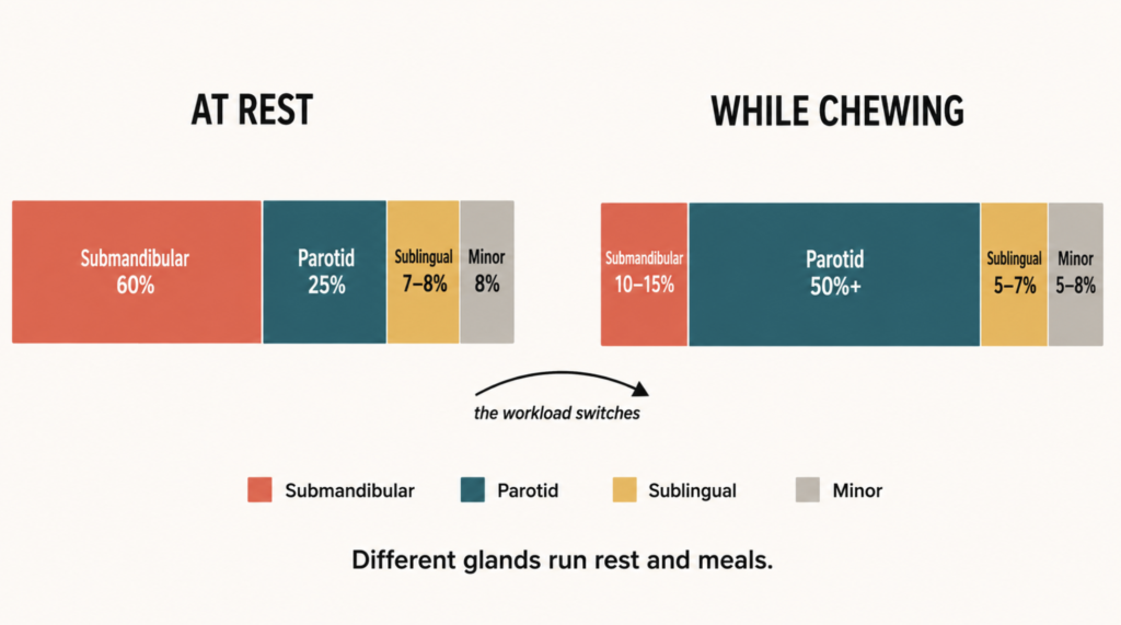
At rest — unstimulated flow:
| Gland | Share of whole saliva |
|---|---|
| Submandibular | ~60% |
| Parotid | ~25% |
| Sublingual | 7–8% |
| Minor glands | ~8% |
During chewing — stimulated flow: the parotid glands take over and account for at least 50% of the total, while every other gland’s percentage share falls [Pedersen, Salivary functions in mastication, taste and textural perception, swallowing and initial digestion, 2018].
The one-line version: the submandibular gland runs your resting mouth; the parotid gland runs your meals.
The stimulus type matters too. Mechanical stimulation — chewing — is heavily parotid-driven. Chemical stimulation from sour taste recruits both parotid and submandibular glands substantially [Pedersen, Salivary functions in mastication, taste and textural perception, swallowing and initial digestion, 2018].
This division has a practical implication worth reasoning through. If the parotid glands supply most of the saliva that arrives during a meal, then damage concentrated in the parotids — as can occur with head and neck radiotherapy fields — would be expected to make eating the hardest part of the day, while resting comfort is comparatively preserved. Reduced submandibular output would be expected to produce the opposite pattern: a constant background dryness largely independent of meals. This is an inference from the physiology rather than a validated diagnostic rule, but it is a reasonable prompt to describe when the dryness is worst, not just that it is there.
Saliva Almost Stops When You Sleep
Here is the number that explains the 4 a.m. glass of water.
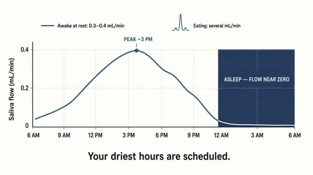
| State | Whole-saliva flow rate |
|---|---|
| Asleep | virtually zero |
| Awake, at rest | 0.3–0.4 mL/min |
| Chewing / eating | several mL/min |
The sleep figure comes from a 1956 study that cannulated the parotid, sublingual, and submaxillary ducts in sleeping subjects and found secretion falling essentially to nothing [Schneyer, Rate of flow of human parotid, sublingual, and submaxillary secretions during sleep, 1956]. It has been reproduced since and remains the reference point [Dawes, Salivary flow patterns and the health of hard and soft oral tissues, 2008].
Salivary flow also follows a genuine circadian rhythm rather than simply tracking activity. When unstimulated whole saliva was sampled repeatedly across the day in the same subjects over weeks, flow rate showed a significant rhythm with its peak in the mid-afternoon [Dawes, Circadian rhythms in human salivary flow rate and composition, 1972]. Your mouth is at its wettest around 3 p.m. and at its driest in the pre-dawn hours. That trough is scheduled.
Two consequences follow.
First, nearly all of the saliva you produce in a day is made while you are awake, and a large share of that during the minutes you spend eating. The seven or eight hours of sleep contribute almost nothing to the daily total.
Second — and this is the part worth acting on — the single most valuable time to brush your teeth is immediately before bed. Saliva provides clearance, buffering, and remineralization. For the entire night, that protective system is offline [Dawes, Salivary flow patterns and the health of hard and soft oral tissues, 2008]. Whatever you leave on your teeth stays there, unbuffered, for eight hours.
But Low Flow Alone Doesn’t Explain Morning Dryness
This is where most explanations stop, and where they go wrong.
Salivary flow collapses overnight in everyone. Yet only some people wake up parched. If reduced secretion were the whole story, morning dry mouth would be universal. It is not. Something else has to account for the difference between the people who wake up fine and the people who keep a bottle on the nightstand.
That something is usually the airway.
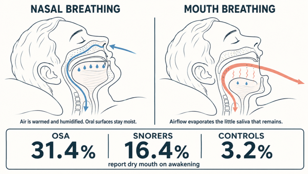
When the mouth hangs open during sleep, air moves continuously across the oral mucosa and evaporates what little saliva is present. The result is a double hit: production has fallen to near zero, and the small residual pool is being actively dried. Nasal breathing does not do this — air is humidified and warmed in the nose and never crosses the oral surfaces.
The epidemiology supports this directly. In a study of 668 adults referred for polysomnography and 582 controls attending a general health check, dry mouth on awakening was reported “almost always” by 31.4% of patients with obstructive sleep apnea, 16.4% of primary snorers, and only 3.2% of controls. The prevalence rose stepwise with disease severity — 22.4% in mild OSA, 34.5% in moderate, 40.7% in severe — and the symptom still distinguished OSA patients from primary snorers after adjusting for age, BMI, sex, hypertension, and other classical OSA symptoms [Oksenberg, Dry mouth upon awakening in obstructive sleep apnea, 2006].
This is cross-sectional data, so it establishes association rather than proving cause. But a roughly tenfold gap between OSA patients and controls, with a dose-response relationship to severity, is difficult to attribute to the salivary glands — every group in that study had the same overnight drop in secretion. The variable that differed was breathing.
Clinical Perspective
When someone says “my mouth is dry when I wake up,” the intuitive move is to think about salivary glands. The evidence above suggests that intuition sends attention to the wrong organ in a meaningful share of cases.
The more useful opening question is not about saliva. It is: do you breathe through your nose when you sleep? Followed by: do you snore, has anyone observed you stopping breathing, are you sleepy during the day, is your nose blocked at night?
A blocked nose forces mouth breathing. Mouth breathing dries the mouth. And chronic mouth breathing during sleep is also how obstructive sleep apnea frequently presents to a clinician who is listening for it. Nasal obstruction from septal deviation, turbinate hypertrophy, or persistent rhinitis is treatable — and treating it addresses the dryness at its source rather than lubricating around it.
Prescribing an artificial saliva spray to a patient whose real problem is that they cannot breathe through their nose is a bandage over a symptom. This is a clinical judgment about where to look first, not a claim that every dry mouth is an airway problem.
When Does Dry Mouth Become a Diagnosis?
Two words get used interchangeably in ordinary speech and mean different things in medicine.
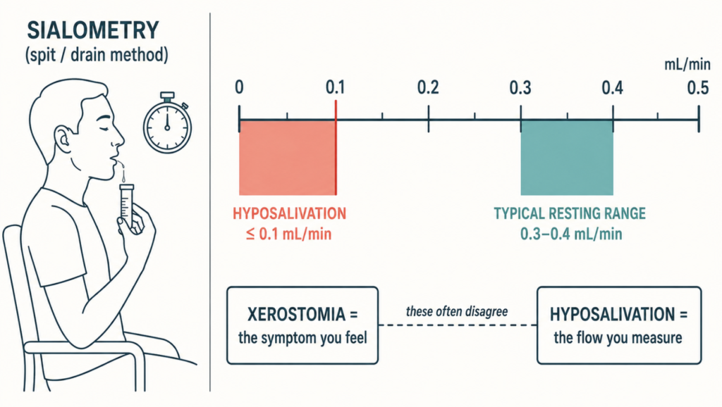
Xerostomia is the subjective complaint — the sensation of a dry mouth. Hyposalivation is the objective finding — a measured reduction in salivary flow.
They correlate imperfectly. Patients report severe dryness with normal measured flow, and patients with clearly reduced flow sometimes report nothing at all [Villa, Diagnosis and management of xerostomia and hyposalivation, 2015]. If you have ever been told your tests came back normal while your mouth still felt like a desert, this distinction is the reason.
The Objective Tests
Sialometry is the practical standard. Unstimulated whole saliva (UWS) is collected by spitting or draining into a container over a timed period; stimulated whole saliva (SWS) is collected while chewing paraffin wax or gum.
The conventional thresholds:
- UWS ≤ 0.1 mL/min — hyposalivation
- SWS ≤ 0.5–0.7 mL/min — hyposalivation
Against a population mean of 0.3–0.4 mL/min at rest, the 0.1 threshold sits at roughly a quarter to a third of typical resting output [Dawes, Salivary flow patterns and the health of hard and soft oral tissues, 2008].
The threshold has a well-known weakness: a meaningful minority of people sit below 0.1 mL/min with no complaint whatsoever. Because individual baselines vary so widely, a 50% drop from a person’s own previous flow rate is often a more meaningful indicator of dysfunction than any absolute cutoff — which is unhelpful in practice, since almost nobody has a baseline measurement on file.
Other tools in use: the modified Schirmer test (a paper strip placed in the mouth), clinical dryness scoring systems, validated questionnaires such as the Xerostomia Inventory, salivary gland ultrasound, scintigraphy, sialography, minor salivary gland biopsy, and anti-SSA/Ro serology when Sjögren syndrome is suspected [Villa, Diagnosis and management of xerostomia and hyposalivation, 2015].
Worth stating plainly: there is no gold standard test for the symptom. There are good tests for the gland.
Who Reports It
The pooled prevalence of dry mouth across 29 population-based studies is 22% (95% CI 17–26%), with clearly higher rates in studies restricted to older people [Agostini, How Common is Dry Mouth? Systematic Review and Meta-Regression Analysis of Prevalence Estimates, 2018]. Estimates in individual studies range widely — from around 10% to over 40% — largely because different studies define and measure the condition differently.
The consistent risk factors:
- Medications — the dominant cause. Hundreds of drugs reduce salivary output, with anticholinergics, antidepressants, antihistamines, antihypertensives, diuretics, and opioids among the most frequent offenders. Risk compounds with the number of drugs taken [Villa, Diagnosis and management of xerostomia and hyposalivation, 2015].
- Head and neck radiotherapy — direct glandular damage, often permanent.
- Sjögren syndrome — autoimmune destruction of exocrine glands; a leading cause of severe, chronic hyposalivation.
- Older age, though see the caveat below.
- Female sex, consistently across studies.
- Diabetes, chronic kidney disease, dehydration, smoking, and alcohol.
One qualification worth making: the common framing that “everyone’s mouth dries out with age” is probably too simple, though the picture is not settled. Studies of healthy older adults generally find that age-related decline in major salivary gland function is modest, while minor gland output falls more clearly — and much of the dry mouth seen in older populations tracks with medication use and accumulated illness rather than with age alone [Villa, Diagnosis and management of xerostomia and hyposalivation, 2015].
The useful reading is that both contribute, and that they are not equally actionable. Some degree of glandular change with age is expected and cannot be reversed; medication burden and comorbidity can often be modified. Assuming a dry mouth is simply “getting older” risks skipping a medication review that might have helped — and assuming it is entirely drug-related risks promising a patient more improvement than adjusting their prescriptions will deliver.
What Actually Helps — And How Much
This is the section most articles handle by listing products. The evidence deserves better than a list.
First: remove the cause
The highest-yield intervention is the one most often skipped. Review the medication list with the prescribing physician for anticholinergic burden and possible substitutions. Restore nasal breathing where obstruction exists. Evaluate and treat obstructive sleep apnea. Check CPAP masks for leak if the dryness began or worsened after starting therapy. Humidify the bedroom.
None of this is glamorous, and all of it addresses mechanism rather than sensation.
Second: topical agents — modest and poorly evidenced
Saliva substitutes, gels, sprays, sugar-free gum, and xylitol lozenges are the usual first line. They are safe and some patients like them. The evidence is weaker than their ubiquity suggests.
A Cochrane review of randomized trials of topical therapies — lozenges, sprays, mouthrinses, gels, oils, chewing gum, toothpastes — concluded that there was no strong evidence that any topical therapy is effective for relieving dry mouth symptoms. Chewing gum reliably increased salivary production, but this did not translate into demonstrated symptom improvement [Furness, Interventions for the management of dry mouth: topical therapies, 2011].
A companion Cochrane review of non-pharmacological approaches, including acupuncture and electrostimulation, reached similarly cautious conclusions on low-quality evidence [Furness, Interventions for the management of dry mouth: non-pharmacological interventions, 2013].
Practical additions worth making regardless: avoid alcohol-containing mouthrinses, which can aggravate the sensation; use fluoride and maintain frequent dental review, because caries prevention is a concrete benefit independent of comfort.
Third: systemic sialogogues — real effect, real cost
Pilocarpine and cevimeline are muscarinic agonists that stimulate residual glandular tissue. Both are approved for dry mouth.
The evidence divides sharply by cause.
In Sjögren syndrome, a meta-analysis of 36 randomized trials including 3,274 patients found high-quality evidence that pilocarpine reduces dry mouth symptoms compared with placebo, and moderate-quality evidence that pilocarpine increases salivary flow — with a large effect size. Rituximab and interferon-alpha also increased flow, with notably smaller effects. Adverse events were common, and the authors concluded that other treatment modalities could not be supported on current evidence [Al Hamad, Interventions for dry mouth and hyposalivation in Sjögren’s syndrome, 2019].
After head and neck radiotherapy, the picture is worse. A Cochrane review of pharmacological prevention found insufficient evidence to determine whether pilocarpine performs better or worse than placebo for xerostomia, salivary flow rate, survival, or quality of life — while finding low-quality evidence that pilocarpine increases sweating [Riley, Pharmacological interventions for preventing dry mouth and salivary gland dysfunction following radiotherapy, 2017].
Two structural limitations apply to this whole drug class. The effect is short-lived, tracking the dosing interval rather than restoring baseline function. And these drugs only work if functional glandular tissue remains — they stimulate, they do not regenerate. Sweating, nausea, urinary frequency, and headache drive substantial discontinuation.
Fourth: prevention and the research frontier
For patients undergoing radiotherapy, the strategy has shifted toward protecting function before it is lost rather than replacing it afterward — parotid-sparing intensity-modulated radiotherapy and surgical submandibular gland transfer are the main approaches, and both fall outside the scope of the drug reviews cited here. On the pharmacological side, radioprotective agents have been studied with mixed results on generally low-quality evidence [Riley, Pharmacological interventions for preventing dry mouth and salivary gland dysfunction following radiotherapy, 2017]. Gene therapy, stem cell approaches, and salivary gland regeneration remain experimental.
The honest summary: current treatment manages the symptom. It does not restore lost function. The largest gains available today come not from a product but from removing the cause — adjusting medications and fixing the airway.
Key Takeaways
- Humans have three pairs of major salivary glands — six in total — plus roughly 600 to 1,000 minor glands scattered through the oral mucosa.
- At rest, about 60% of saliva comes from the submandibular glands; during chewing, the parotid glands supply at least 50%.
- Salivary flow falls to virtually zero during sleep, compared with 0.3–0.4 mL/min while awake and at rest.
- Because overnight flow drops in everyone, persistent morning dryness usually points to mouth breathing, snoring, or obstructive sleep apnea rather than to the salivary glands themselves.
- Xerostomia is the subjective symptom; hyposalivation is the measured sign. They frequently disagree, which is why normal test results do not invalidate a real complaint.
- Dry mouth affects around 22% of adults; age and medication use both contribute, but medication burden is the more modifiable of the two.
- Cochrane reviews found no strong evidence that any topical therapy relieves dry mouth symptoms, though chewing gum does increase saliva production.
- Pilocarpine has high-quality evidence for symptom relief in Sjögren syndrome but adverse events are common, and evidence after radiotherapy is insufficient.
FAQ
How many salivary glands do humans have? Six major glands and several hundred minor ones. The major glands come in three pairs — parotid, submandibular, and sublingual — and produce about 90% of total salivary volume. The 600 to 1,000 minor glands in the lips, cheeks, palate, and tongue supply the remaining 10%, but contribute a disproportionate share of the lubricating mucins.
Does saliva completely stop during sleep? Effectively, yes. Direct measurements of the major glands during sleep found flow falling to virtually zero, against a waking resting rate of 0.3–0.4 mL/min. Salivary flow follows a circadian rhythm that peaks in the mid-afternoon and bottoms out overnight, so the drop is scheduled rather than incidental.
Is waking up with a dry mouth normal? Occasionally, yes — overnight flow drops in everyone. Daily, severe morning dryness is different. Because the reduction in secretion is universal, the people who wake up parched usually have an additional factor, most often mouth breathing from nasal obstruction, snoring, or sleep apnea.
What salivary flow rate is considered too low? Unstimulated whole saliva at or below 0.1 mL/min, and stimulated whole saliva at or below roughly 0.5–0.7 mL/min, are the conventional thresholds for hyposalivation. These cutoffs have limits: individual baselines vary enormously, and some people sit below the threshold without any symptoms.
Can morning dry mouth be a sign of sleep apnea? It can be. In one study, dry mouth on awakening was reported by 31.4% of patients with obstructive sleep apnea versus 3.2% of controls, and the rate rose with apnea severity. Morning dryness combined with loud snoring or daytime sleepiness is worth raising with a physician.
Do artificial saliva products actually work? The evidence is weaker than their availability suggests. A Cochrane review found no strong evidence that any topical therapy — sprays, gels, rinses, lozenges, gum — improves dry mouth symptoms, though chewing gum does increase saliva production. They are safe and some patients find them helpful, so preference and tolerability reasonably guide the choice.
Does dry mouth mean I have Sjögren syndrome? Usually not. Medications are a far more common cause. Sjögren syndrome is worth investigating when dry mouth occurs alongside dry eyes, salivary gland swelling, joint symptoms, or marked fatigue, and diagnosis relies on a combination of symptoms, salivary flow measurement, autoantibody testing, and sometimes minor salivary gland biopsy.
References
- Agostini BA, Cericato GO, Silveira ERD, Nascimento GG, Costa FDS, Thomson WM, Demarco FF. How Common is Dry Mouth? Systematic Review and Meta-Regression Analysis of Prevalence Estimates. Braz Dent J. 2018 Nov-Dec;29(6):606-618.
- Al Hamad A, Lodi G, Porter S, Fedele S, Mercadante V. Interventions for dry mouth and hyposalivation in Sjögren’s syndrome: A systematic review and meta-analysis. Oral Dis. 2019 May;25(4):1027-1047.
- Dawes C. Circadian rhythms in human salivary flow rate and composition. J Physiol. 1972 Feb;220(3):529-545.
- Dawes C. Salivary flow patterns and the health of hard and soft oral tissues. J Am Dent Assoc. 2008 May;139 Suppl:18S-24S.
- Furness S, Worthington HV, Bryan G, Birchenough S, McMillan R. Interventions for the management of dry mouth: topical therapies. Cochrane Database Syst Rev. 2011 Dec 7;(12):CD008934.
- Furness S, Bryan G, McMillan R, Birchenough S, Worthington HV. Interventions for the management of dry mouth: non-pharmacological interventions. Cochrane Database Syst Rev. 2013 Sep 5;(9):CD009603.
- Oksenberg A, Froom P, Melamed S. Dry mouth upon awakening in obstructive sleep apnea. J Sleep Res. 2006 Sep;15(3):317-320.
- Pedersen AML, Sørensen CE, Proctor GB, Carpenter GH. Salivary functions in mastication, taste and textural perception, swallowing and initial digestion. Oral Dis. 2018 Nov;24(8):1399-1416.
- Riley P, Glenny AM, Hua F, Worthington HV. Pharmacological interventions for preventing dry mouth and salivary gland dysfunction following radiotherapy. Cochrane Database Syst Rev. 2017 Jul 31;7(7):CD012744.
- Schneyer LH, Pigman W, Hanahan L, Gilmore RW. Rate of flow of human parotid, sublingual, and submaxillary secretions during sleep. J Dent Res. 1956 Feb;35(1):109-114.
- Villa A, Connell CL, Abati S. Diagnosis and management of xerostomia and hyposalivation. Ther Clin Risk Manag. 2015;11:45-51.
Joonpyo Hong, MD is a board-certified otolaryngologist practicing in Korea. This article reflects his clinical interpretation of published research and does not constitute individual medical advice.
For more interesting content:
https://curiousmd.com/salivary-gland-stones-treatment/
https://curiousmd.com/facial-and-neck-lacerations-by-site/
Link out to:
Dry Mouth — National Institute of Dental and Craniofacial Research (NIDCR) — the NIH institute responsible for salivary research; states plainly that dry mouth is not a normal part of aging.
Dry Mouth — MedlinePlus, U.S. National Library of Medicine — patient-level overview of causes, diagnosis, and self-care.
Oksenberg A, et al. Dry mouth upon awakening in obstructive sleep apnea. J Sleep Res. 2006 — PubMed — the case-control study behind the 31.4% vs 3.2% figures cited above.
Agostini BA, et al. How Common is Dry Mouth? Braz Dent J. 2018 — PubMed — the meta-analysis behind the 22% pooled prevalence estimate.