
If a dentist has ever pointed out fine white lines inside your cheek, you may have met oral lichen planus. It is an immune condition that affects about 1% of people worldwide [González-Moles, Worldwide prevalence of oral lichen planus: A systematic review and meta-analysis, 2021], and in one large clinic series almost half of patients had no symptoms at all [Budimir, Oral lichen planus – retrospective study of 563 Croatian patients, 2014]. Online, you’ll read that it is harmless, and also that it is precancerous. There is some truth in both.
What oral lichen planus is and why it happens
Oral lichen planus is a long-lasting inflammatory condition in which T cells, a type of immune cell, attack the bottom layer of cells lining the mouth [Akhtar, Oral Lichen Planus, 2025]. Think of security guards who mistake the building’s own staff for intruders. The staff at the base of the lining get damaged, and the white lines are the surface thickening in response.
What makes the guards misfire isn’t known. Reported associations include certain medicines, dental materials, hepatitis C, stress, and friction from sharp teeth or rough fillings. Hepatitis C is more common in people with lichen planus, though the link varies between studies [Lodi, Hepatitis C virus infection and lichen planus: a systematic review with meta-analysis, 2010]. Oral lichen planus is not an infection, and it can’t be passed to anyone [American Academy of Oral Medicine, Oral Lichen Planus, 2013].
Lichen planus vs. oral lichenoid lesions
Some look-alike lesions are reactions to a medicine or a dental material, often an amalgam filling that touches the lesion. Drug reactions are more often one-sided [Gururaj, Diagnosis and management of oral lichen planus – Review, 2021]. Once the trigger is removed, these can clear within months.
Where it shows up: cheeks, tongue, gums, and elsewhere
The inner cheek is the classic site. In the Croatian series it was involved in 82.4% of patients, usually on both sides, followed by the tongue, gums, and lips. About 10% of patients have disease only on the gums, which look red and peel; this pattern is called desquamative gingivitis [Akhtar, Oral Lichen Planus, 2025].
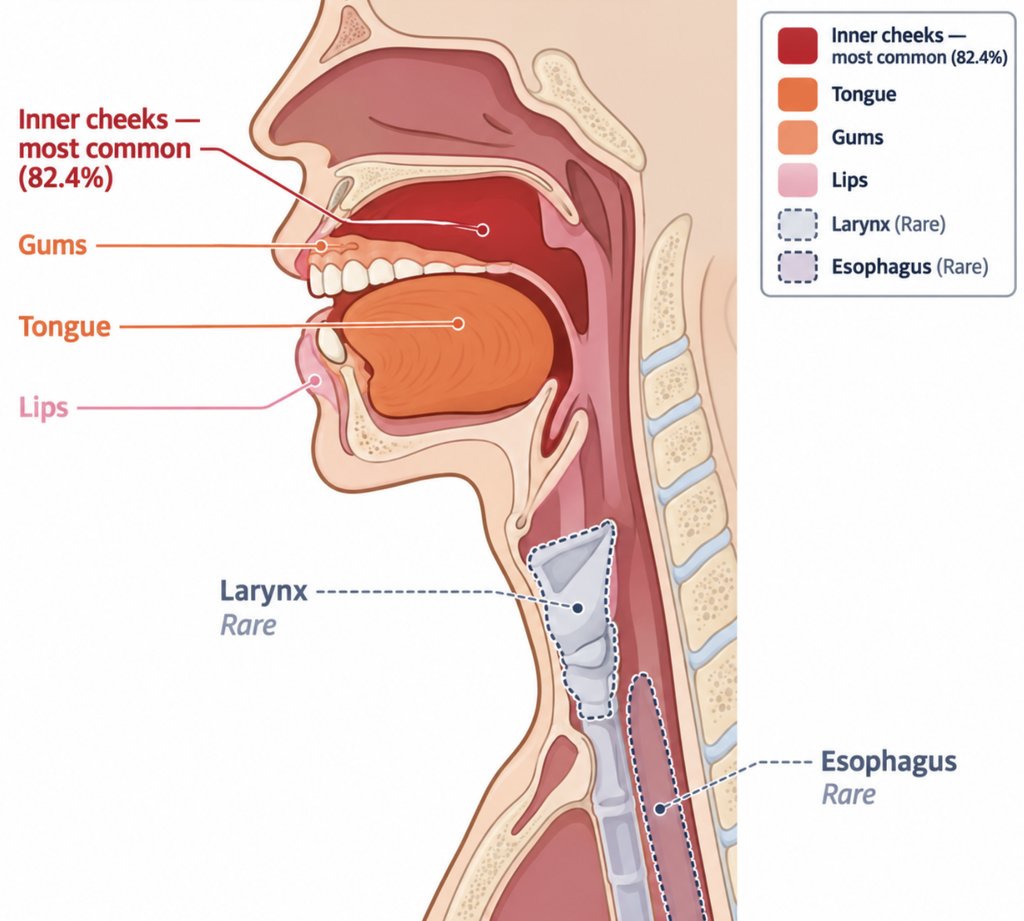
Outside the mouth, about 15% of people with the oral form develop skin lesions, and around 20% of women develop genital lesions. The oesophagus can also be involved, which may make swallowing difficult or painful. Involvement of the larynx is limited to a handful of reported cases, some with hoarseness [Gorouhi, Cutaneous and mucosal lichen planus: a comprehensive review of clinical subtypes, risk factors, diagnosis, and prognosis, 2014]. If you already have oral lichen planus, new trouble swallowing or a voice change that doesn’t go away is worth an endoscopic look.
The six forms and how they look and feel
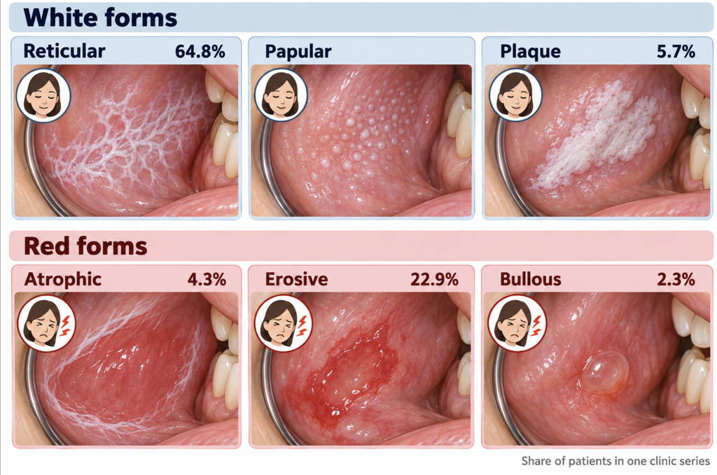
Oral lichen planus is described in six clinical forms, grouped as white or red [Akhtar, Oral Lichen Planus, 2025].
| Form | Appearance | Typical symptoms | Share in one series |
|---|---|---|---|
| Reticular (white) | Lacy white lines (Wickham striae) | Usually none | 64.8% |
| Papular (white) | Tiny white dots | Often painless | Not reported |
| Plaque (white) | Thick white patch | Often painless | 5.7% |
| Atrophic (red) | Thin, glazed red lining | Burning with spicy food | 4.3% |
| Erosive (red) | Raw areas or open sores | Pain eating or brushing | 22.9% |
| Bullous (red) | Blisters that break | Pain | 2.3% |
Percentages come from the Croatian series.
Reticular is the most common form and usually causes no symptoms; in the Croatian series, 44.2% of patients had none. The red forms are the ones that hurt, especially with spicy or acidic food.
Does oral lichen planus need a biopsy?
A biopsy confirms the diagnosis under the microscope. Whether every lacy white patch needs one at the first visit is where experts disagree. The American Academy of Oral and Maxillofacial Pathology calls for a biopsy to make a definitive diagnosis [Cheng, Diagnosis of oral lichen planus: a position paper of the American Academy of Oral and Maxillofacial Pathology, 2016]. Other reviewers accept that the classic reticular form can often be diagnosed by its appearance, and use biopsy mainly to look for dysplasia, the abnormal cell changes that come before cancer [Akhtar, Oral Lichen Planus, 2025].
The risk isn’t spread evenly. In a large meta-analysis, atrophic or erosive lesions carried about four times the cancer risk, and tongue lesions nearly twice [González-Moles, Malignant transformation risk of oral lichen planus: A systematic review and comprehensive meta-analysis, 2019]. Specialists generally favor a biopsy when a lesion shows:
- No lacy white lines anywhere, since that pattern is the hallmark of the disease.
- One side only, or contact with a filling, which suggests a lichenoid reaction.
- A thick white plaque, or a patchy mix of red and white.
- A red or eroded area, especially one that doesn’t heal with treatment, or an area that feels firm.
- A site on the side of the tongue or the floor of the mouth.
- Any change between check-ups.
Cancer estimates range from 0.44% under strict diagnostic criteria [Idrees, Oral lichen planus has a very low malignant transformation rate, 2021] to 2.28% in the highest-quality follow-up studies [González-Moles, An appraisal of highest quality studies reporting malignant transformation of oral lichen planus based on a systematic review, 2021]. Dysplasia is what a biopsy looks for, and it changes the odds: lesions with dysplasia have been estimated to become cancer in around 6% of cases, compared with under 1.5% without it [González-Moles, Dysplasia in oral lichen planus: relevance, controversies and challenges, 2021].
In a Finnish cohort, patients who developed cancer did so an average of 11.4 years after diagnosis [Vehviläinen, Risk of Malignant Transformation in Patients With Oral Lichen Planus or Lichenoid Lesions, 2026], so follow-up continues even when nothing seems to change.
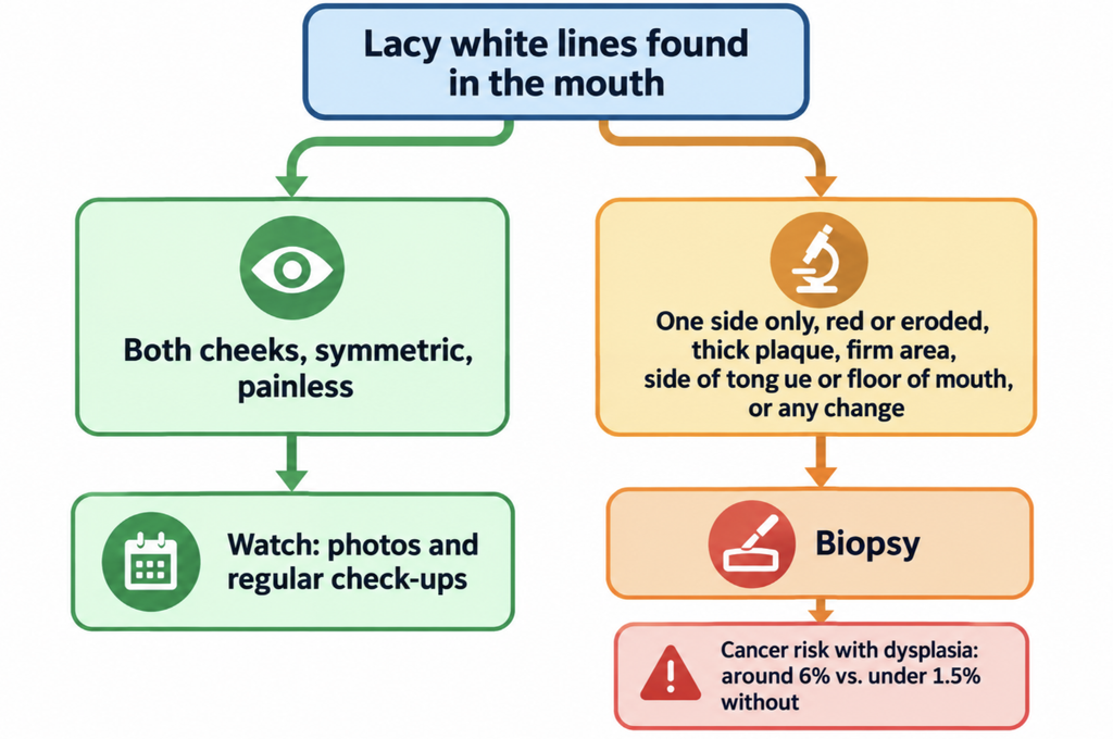
Clinical Perspective. A painless lacy pattern on both cheeks is usually observed and photographed rather than biopsied. The plan changes if the lesion becomes asymmetric, if a red or ulcerated area doesn’t respond to treatment, if it thickens, or if something looks different at the next visit. An otolaryngologist can check the throat and larynx at the same visit.
Who gets oral lichen planus?
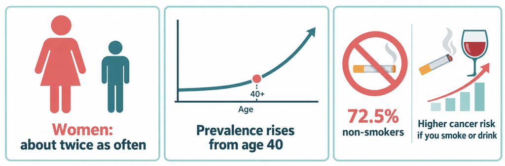
Oral lichen planus mainly affects adults, and prevalence climbs from age 40 onward. In the Croatian series, the average age at diagnosis was 58. Women get the oral form about twice as often as men, while the skin form affects both sexes roughly equally [Johns Hopkins Medicine, Lichen Planus, 2019]. It is rare in children, at about 0.03% [Spirito, Oral lichen planus in children: A systematic review, 2024].
Smoking and alcohol
Oral lichen planus is not a smoker’s disease. In the Croatian series, 72.5% of patients did not smoke, and neither smoking nor drinking has been shown to cause the condition.
Once someone has it, though, both matter. In pooled data, smokers with oral lichen planus had about twice the cancer risk, and drinkers slightly more than twice [González-Moles, Malignant transformation risk of oral lichen planus: A systematic review and comprehensive meta-analysis, 2019]. Tobacco and alcohol do not light the fire, but they add fuel.
Treatment and follow-up
There is no cure. Painless reticular lesions often need nothing more than periodic checks. For painful lesions, corticosteroid gels or rinses are the usual first step. A Cochrane review found they may relieve pain better than placebo, though the evidence is limited [Lodi, Interventions for treating oral lichen planus: corticosteroid therapies, 2020]. Oral thrush is a common side effect.
Other common advice: have sharp teeth or rough fillings smoothed, avoid spicy food, use a mild toothpaste, and stop tobacco and alcohol. Keep your follow-up appointments, even when nothing hurts.
Key Takeaways
- Oral lichen planus is a chronic, non-contagious immune condition that affects about 1% of people.
- The classic form is a painless, lacy white pattern on both inner cheeks.
- A classic two-sided pattern can often be watched. Red, ulcerated, thick, one-sided, or changing lesions deserve a biopsy.
- Cancer risk is low, estimated at 0.44% to 2.28%, and higher when dysplasia is present.
- It mostly affects women over 40, many of whom don’t smoke. In people who have it, smoking and alcohol raise the cancer risk.
FAQ
Is oral lichen planus contagious?
No. It is an immune reaction, not an infection, so it can’t be caught or passed on [American Academy of Oral Medicine, Oral Lichen Planus, 2013].
Can oral lichen planus turn into cancer?
Rarely. Estimates range from 0.44% to 2.28%, with higher risk in red or ulcerated forms and in lesions with dysplasia [González-Moles, Malignant transformation risk of oral lichen planus: A systematic review and comprehensive meta-analysis, 2019].
Does oral lichen planus go away?
Usually not. It flares and settles over years, and treatment aims to keep it painless rather than make it disappear [American Academy of Oral Medicine, Oral Lichen Planus, 2013].
Can stress trigger oral lichen planus?
Possibly. Flare-ups often coincide with stressful periods, although stress has not been shown to cause the condition on its own [Akhtar, Oral Lichen Planus, 2025].
References
- González-Moles MÁ, Warnakulasuriya S, González-Ruiz I, González-Ruiz L, Ayén Á, Lenouvel D, et al. Worldwide prevalence of oral lichen planus: A systematic review and meta-analysis. Oral Dis. 2021 May;27(4):813-28.
- Budimir V, Richter I, Andabak-Rogulj A, Vučićević-Boras V, Budimir J, Brailo V. Oral lichen planus – retrospective study of 563 Croatian patients. Med Oral Patol Oral Cir Bucal. 2014 May 1;19(3):e255-60.
- Akhtar N, Brizuela M. Oral Lichen Planus. In: StatPearls [Internet]. Treasure Island (FL): StatPearls Publishing; 2025 [updated 2025 Sep 15].
- Lodi G, Pellicano R, Carrozzo M. Hepatitis C virus infection and lichen planus: a systematic review with meta-analysis. Oral Dis. 2010 Oct;16(7):601-12.
- American Academy of Oral Medicine. Oral Lichen Planus [Internet]. Atlanta (GA): AAOM; 2013 [cited 2026 Sep 27]. Available from: https://www.aaom.com/oral-lichen-planus
- Gururaj N, Hasinidevi P, Janani V, Divynadaniel T. Diagnosis and management of oral lichen planus – Review. J Oral Maxillofac Pathol. 2021;25(3):383-93.
- Gorouhi F, Davari P, Fazel N. Cutaneous and mucosal lichen planus: a comprehensive review of clinical subtypes, risk factors, diagnosis, and prognosis. ScientificWorldJournal. 2014;2014:742826.
- Cheng YS, Gould A, Kurago Z, Fantasia J, Muller S. Diagnosis of oral lichen planus: a position paper of the American Academy of Oral and Maxillofacial Pathology. Oral Surg Oral Med Oral Pathol Oral Radiol. 2016 Sep;122(3):332-54.
- González-Moles MÁ, Ruiz-Ávila I, González-Ruiz L, Ayén Á, Gil-Montoya JA, Ramos-García P. Malignant transformation risk of oral lichen planus: A systematic review and comprehensive meta-analysis. Oral Oncol. 2019 Sep;96:121-30.
- Vehviläinen M, Selander T, Salo T, Siponen M. Risk of Malignant Transformation in Patients With Oral Lichen Planus or Lichenoid Lesions: A Retrospective Study of 323 Patients With a Minimum of 5-Year Follow-Up. J Oral Pathol Med. 2026;55(7):784-92.
- Idrees M, Kujan O, Shearston K, Farah CS. Oral lichen planus has a very low malignant transformation rate: A systematic review and meta-analysis using strict diagnostic and inclusion criteria. J Oral Pathol Med. 2021 Mar;50(3):287-98.
- González-Moles MÁ, Warnakulasuriya S, González-Ruiz I, Ayén Á, González-Ruiz L, Ruiz-Ávila I, et al. Dysplasia in oral lichen planus: relevance, controversies and challenges. A position paper. Med Oral Patol Oral Cir Bucal. 2021 Jul 1;26(4):e541-8.
- Johns Hopkins Medicine. Lichen Planus [Internet]. Baltimore (MD): Johns Hopkins Medicine; 2019 [cited 2026 Sep 27]. Available from: https://www.hopkinsmedicine.org/health/conditions-and-diseases/lichen-planus
- Spirito F, Dioguardi M, Caponio VC, Ambrosino M, Lo Muzio E, Lo Muzio L. Oral lichen planus in children: A systematic review. Med Oral Patol Oral Cir Bucal. 2024 Mar 1;29(2):e152-62.
- Lodi G, Manfredi M, Mercadante V, Murphy R, Carrozzo M. Interventions for treating oral lichen planus: corticosteroid therapies. Cochrane Database Syst Rev. 2020 Feb 28;2(2):CD001168.
- González-Moles MÁ, Ramos-García P, Warnakulasuriya S. An appraisal of highest quality studies reporting malignant transformation of oral lichen planus based on a systematic review. Oral Dis. 2021 Nov;27(8):1908-18.
Joonpyo Hong, MD is a board-certified otolaryngologist practicing in Korea. This article reflects his clinical interpretation of published research and does not constitute individual medical advice.
For more articles:
https://curiousmd.com/oral-cancer-swab-test-accuracy-explained/
https://curiousmd.com/tooth-infection-spread-to-neck-timeline/
https://curiousmd.com/sore-throat-on-one-side-peritonsillar-abscess/
Linkt out to:
https://www.aaom.com/oral-lichen-planus — The American Academy of Oral Medicine’s patient guide to oral lichen planus patterns, biopsy, treatment, and cancer questions.
https://www.hopkinsmedicine.org/health/conditions-and-diseases/lichen-planus — Johns Hopkins Medicine’s overview of skin and oral lichen planus, who gets it, and how it is managed.
https://my.clevelandclinic.org/health/diseases/17875-oral-lichen-planus — Cleveland Clinic’s patient guide to oral lichen planus symptoms, triggers, and treatment.
https://cancer.ca/en/cancer-information/cancer-types/oral/risks — The Canadian Cancer Society’s summary of oral cancer risk factors, including long-standing ulcerative lichen planus.