
A Tornwaldt cyst on MRI usually shows up as a single line in a report for a scan ordered for something else: headaches, dizziness, a neck problem. The phrase “cyst in the nasopharynx” can sound alarming, because many people link a lump behind the nose with cancer. Below: what the cyst is, how often scans find it by chance, and when it needs treatment.
What Is a Tornwaldt Cyst?
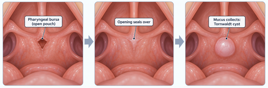
A Tornwaldt cyst (also spelled Thornwaldt cyst) is a benign, fluid-filled cyst in the midline of the back wall of the nasopharynx, the air space behind the nose and above the soft palate [Cetinkaya, Thornwaldt Cyst, 2018]. In the embryo, a structure called the notochord touches the lining of the throat and leaves a small pouch there, the pharyngeal bursa. If the pouch’s opening seals over, mucus collects inside and forms a cyst [Cetinkaya, Thornwaldt Cyst, 2018].
Picture a pocket left over from construction. It causes no trouble while it stays open; you notice it only after it seals and fills. Most of these cysts are small. In one MRI series, the average cyst measured about 6 mm across [Ikushima, MR imaging of Tornwaldt’s cysts, 1999].
How Often Is It Found by Chance on CT or MRI?
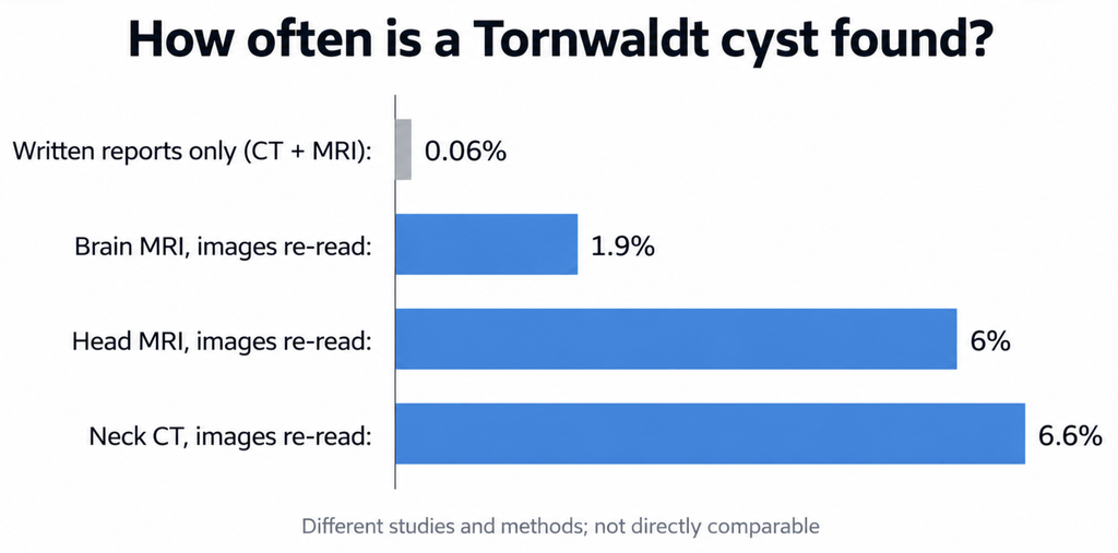
Researchers who re-read routine head MRI scans found a Tornwaldt cyst in roughly 1.9% to 6% of people [Ikushima, MR imaging of Tornwaldt’s cysts, 1999; Sekiya, Nasopharyngeal cystic lesions: Tornwaldt and mucous retention cysts of the nasopharynx, 2014]. That works out to between about 1 in 50 and 1 in 17 scans, so radiologists treat it as a common incidental finding. Patients in these studies had their scans for unrelated reasons, much like a screening brain MRI.
Rare or common? Why sources disagree
Some references call the cyst rare, while radiology texts call it common. The answer depends on how researchers count.
| Study | Scans reviewed | How cysts were counted | Rate found |
|---|---|---|---|
| Moody et al. | 53,013 CT and MRI reports | Cysts mentioned in written reports | 0.06% |
| Ikushima et al. | 1,208 brain MRI scans | Images re-reviewed by researchers | 1.9% |
| Sekiya et al. | 3,000 head MRI scans | Images re-reviewed by researchers | 6% |
| Beheshtian et al. | 2,100 cervical spine CT scans | Images re-reviewed by researchers | 6.6% |
Researchers who searched only written reports found a rate of about 6 in 10,000 [Moody, Tornwaldt’s cyst: incidence and a case report, 2007]. A likely explanation: radiologists often leave small, harmless findings out of the report. In the cervical spine CT study, official reports mentioned only 2.9% of incidental findings of any kind [Beheshtian, Incidental findings on cervical spine computed tomography scans: overlooked and unimportant?, 2018].
In the largest MRI series, cysts peaked at ages 51–60, and the authors suggested that cysts may form and later shrink over a lifetime [Sekiya, Nasopharyngeal cystic lesions: Tornwaldt and mucous retention cysts of the nasopharynx, 2014].
Why MRI catches it more often than CT
A Tornwaldt cyst appears bright on T2-weighted MRI images and often on T1-weighted images too, depending on the protein content of its fluid [Ikushima, MR imaging of Tornwaldt’s cysts, 1999] [Radiopaedia, Tornwaldt cyst]. CT detects and characterizes these cysts less well than MRI [Cetinkaya, Thornwaldt Cyst, 2018]. In the report-based study, radiologists mentioned cysts in 0.13% of MRI reports and 0.013% of CT reports, a tenfold difference, though the two patient groups were not directly comparable [Moody, Tornwaldt’s cyst: incidence and a case report, 2007].
Found a Tornwaldt cyst on MRI: Treat or Watch?
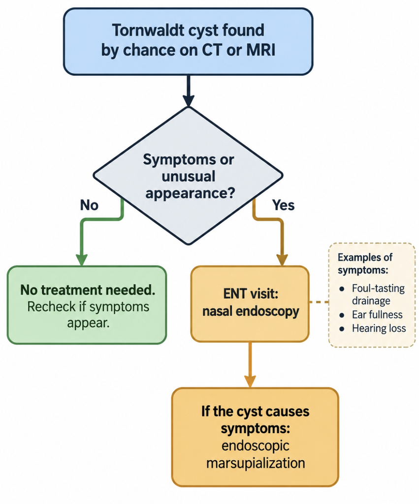
A Tornwaldt cyst that causes no symptoms needs no treatment. Reviews and clinical references agree on this [Cetinkaya, Thornwaldt Cyst, 2018; Cheng, Tornwaldt Cyst (Merck Manual), 2026].
| Situation | Usual approach |
|---|---|
| Found by chance, no symptoms, typical appearance | No treatment needed |
| Persistent foul-tasting drainage down the throat, bad breath, or headache at the back of the head | See an ENT specialist for nasal endoscopy |
| Ear fullness, hearing loss, or fluid behind the eardrum | ENT evaluation; a large cyst can block the Eustachian tube |
| Larger cyst, roughly over 1 to 2 cm | More likely to cause symptoms; ENT review is reasonable |
| Unusual appearance (thick wall, enhancement with contrast) | Endoscopy, and biopsy if needed, to rule out other lesions |
The size threshold comes from case reports and serves as a rough guide [Lee, Huge Tornwaldt Cyst With Otitis Media With Effusion, 2021].
Do you need a follow-up scan?
No formal guideline sets a follow-up schedule for an incidental Tornwaldt cyst, and none of the reviews and references consulted here call for routine repeat imaging. In practice, doctors look again when symptoms change. You and your treating doctor can make that call together.
When treatment is needed
For a cyst that causes symptoms, surgeons usually perform endoscopic marsupialization through the nose: they open the roof of the cyst so it drains and does not refill [Cetinkaya, Thornwaldt Cyst, 2018]. A small series followed 11 patients for 5 years after this procedure and recorded no recurrences or complications [El-Anwar, 5 years follow up after transnasal endoscopic surgery of Thornwaldt’s cyst with powered instrumentation, 2015]. Larger studies are lacking.
Symptoms That Change the Plan
An inflamed or infected cyst, a condition sometimes called Tornwaldt disease, can cause persistent pus-like drainage with a foul taste or smell, a sore throat, and blockage of the Eustachian tube [Cheng, Tornwaldt Cyst (Merck Manual), 2026]. Large cysts can also cause nasal obstruction, snoring, bad breath, and one-sided hearing loss [Bozan, A huge Thornwaldt’s cyst causing hearing loss in an adult patient, 2017].
None of these symptoms point to the cyst alone. In one MRI series, 3 of 23 people with cysts reported nasal discharge or headaches at the back of the head, but the study design could not show whether the cyst caused them [Ikushima, MR imaging of Tornwaldt’s cysts, 1999]. Sinusitis, allergies, and tension headaches produce the same complaints and are far more common.
Could It Be Something Else?
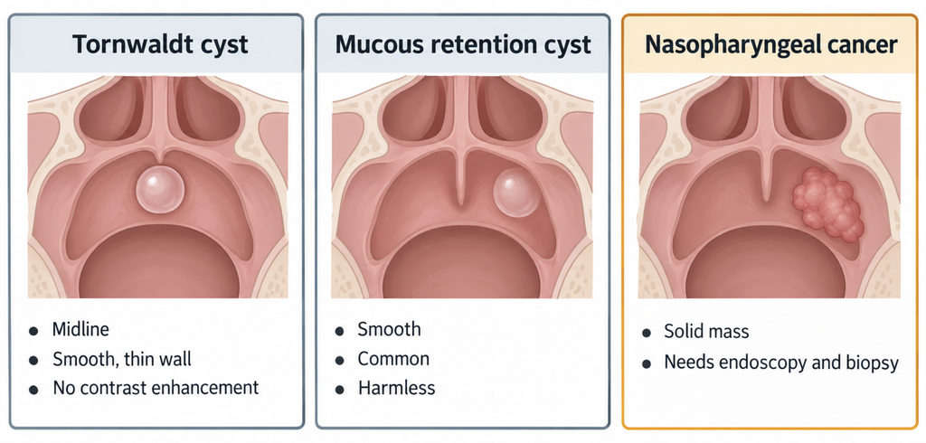
A typical Tornwaldt cyst looks smooth, round, and thin-walled, sits in the midline, and does not light up with contrast dye [Radiopaedia, Tornwaldt cyst]. Its most common look-alike is the mucous retention cyst. In the largest MRI series, mucous retention cysts turned up in about 1 in 10 scans, more often than Tornwaldt cysts [Sekiya, Nasopharyngeal cystic lesions: Tornwaldt and mucous retention cysts of the nasopharynx, 2014].
Nasopharyngeal cancer usually forms a solid mass rather than a fluid-filled sac. Doctors diagnose it by looking at the area through a thin scope and taking a biopsy, often alongside MRI [National Cancer Institute, Nasopharyngeal Cancer Treatment (PDQ)–Patient Version]. Warning signs include a neck lump, nosebleeds, ear pain or ringing, trouble hearing, and a stuffy nose [National Cancer Institute, Nasopharyngeal Cancer Treatment (PDQ)–Patient Version].
Key Takeaways
- A Tornwaldt cyst is a benign, fluid-filled cyst left over from fetal development, located in the midline behind the nose.
- Studies that re-read routine head MRI scans find it in roughly 1.9% to 6% of people.
- An asymptomatic Tornwaldt cyst needs no treatment, and no guideline requires routine follow-up scans.
- Persistent foul-tasting drainage, ear blockage, or an unusual imaging appearance are reasons to see an ENT specialist.
FAQ
Is a Tornwaldt cyst dangerous?
No. It is a benign developmental cyst, and most cause no symptoms [Cetinkaya, Thornwaldt Cyst, 2018]. Problems arise mainly if it becomes infected or grows large enough to block the Eustachian tube [Cheng, Tornwaldt Cyst (Merck Manual), 2026].
How common is a Tornwaldt cyst on MRI?
Fairly common. Studies that re-read routine head MRI scans found it in about 1.9% to 6% of people [Ikushima, MR imaging of Tornwaldt’s cysts, 1999; Sekiya, Nasopharyngeal cystic lesions: Tornwaldt and mucous retention cysts of the nasopharynx, 2014].
Does a Tornwaldt cyst need surgery?
Usually not. Asymptomatic cysts need no treatment [Righi, Thornwaldt cysts, 2014]. Surgeons reserve surgery, usually endoscopic marsupialization through the nose, for cysts that cause symptoms [Cetinkaya, Thornwaldt Cyst, 2018].
Do I need a follow-up scan?
Not routinely. No formal guideline sets a schedule for repeat imaging of an incidental, symptom-free cyst. New symptoms such as persistent drainage or ear fullness are the usual reason to recheck [Cheng, Tornwaldt Cyst (Merck Manual), 2026].
References
- Cetinkaya EA. Thornwaldt Cyst. J Craniofac Surg. 2018;29(6):e560-e562.
- Ikushima I, Korogi Y, Makita O, Komohara Y, Kawano H, Yamura M, et al. MR imaging of Tornwaldt’s cysts. AJR Am J Roentgenol. 1999;172(6):1663-5.
- Sekiya K, Watanabe M, Nadgir RN, Buch K, Flower EN, Kaneda T, et al. Nasopharyngeal cystic lesions: Tornwaldt and mucous retention cysts of the nasopharynx: findings on MR imaging. J Comput Assist Tomogr. 2014;38(1):9-13.
- Moody MW, Chi DH, Mason JC, Phillips CD, Gross CW, Schlosser RJ. Tornwaldt’s cyst: incidence and a case report. Ear Nose Throat J. 2007;86(1):45-7, 52.
- Beheshtian E, Sahraian S, Yousem DM, Khan MK. Incidental findings on cervical spine computed tomography scans: overlooked and unimportant? Neuroradiology. 2018;60(11):1175-1180.
- El-Anwar MW, Amer HS, Elnashar I, Askar SM, Ahmed AF. 5 years follow up after transnasal endoscopic surgery of Thornwaldt’s cyst with powered instrumentation. Auris Nasus Larynx. 2015;42(1):29-33.
- Lee JH. Huge Tornwaldt Cyst With Otitis Media With Effusion. Ear Nose Throat J. 2021;100(5_suppl):528S-530S.
- Righi S, Boffano P, Pateras D, Malvè L, Chiodo D, Boson M. Thornwaldt cysts. J Craniofac Surg. 2014;25(5):e456-7.
- Bozan N, Sakin YF, Kundi P, Ari M, Bozkus F. A huge Thornwaldt’s cyst causing hearing loss in an adult patient. J Pak Med Assoc. 2017;67(3):468-470.
- Cheng AG. Tornwaldt Cyst. Merck Manual Professional Version [Internet]. Rahway (NJ): Merck & Co.; 2026 [cited 2026 Oct 9]. Available from: https://www.merckmanuals.com/professional/ear-nose-and-throat-disorders/oral-and-pharyngeal-disorders/tornwaldt-cyst
- Radiopaedia. Tornwaldt cyst [Internet]. [cited 2026 Oct 9]. Available from: https://radiopaedia.org/articles/tornwaldt-cyst
- National Cancer Institute. Nasopharyngeal Cancer Treatment (PDQ)–Patient Version [Internet]. Bethesda (MD): National Cancer Institute; [cited 2026 Oct 9]. Available from: https://www.cancer.gov/types/head-and-neck/patient/adult/nasopharyngeal-treatment-pdq
Joonpyo Hong, MD is a board-certified otolaryngologist practicing in Korea. This article reflects his clinical interpretation of published research and does not constitute individual medical advice.
For more articles:
https://curiousmd.com/nasopharyngeal-carcinoma-ai-detection/
https://curiousmd.com/ai-virtual-contrast-enhancement/
Link out to:
https://radiopaedia.org/articles/tornwaldt-cyst — Radiopaedia’s radiology reference on Tornwaldt cyst, covering typical CT and MRI appearance and treatment.
https://www.merckmanuals.com/professional/ear-nose-and-throat-disorders/oral-and-pharyngeal-disorders/tornwaldt-cyst — Merck Manual Professional overview of Tornwaldt cyst diagnosis and management (updated June 2026).
https://www.cancer.gov/types/head-and-neck/patient/adult/nasopharyngeal-treatment-pdq — National Cancer Institute patient guide to nasopharyngeal cancer symptoms and diagnostic tests.
https://www.acr.org/Clinical-Resources/Clinical-Tools-and-Reference/Incidental-Findings — American College of Radiology resources on managing findings discovered on scans done for other reasons.