
Ultrasound for nasal bone fracture is usually discussed as a possible substitute for CT. The published evidence does not settle that comparison, and framing it that way skips past the uses the data actually supports.
Take a hypothetical nine-year-old who arrives two hours after a playground collision, nose swollen, dorsum slightly off axis. The usual next step is an X-ray or a CT scan. A three-minute bedside scan is a third option, and on the published evidence it outperforms one of the other two.
The nasal bone is the most frequently fractured bone in the face [Jeong Y, Development of AI-Based Diagnostic Algorithm for Nasal Bone Fracture Using Deep Learning, 2024], so this is one of the most repeated imaging decisions in an otolaryngology clinic.
How ultrasound sees a broken nose
The nasal bone is an unusually good ultrasound target. It sits under thin skin with almost no intervening soft tissue, so a high-frequency linear transducer images the cortical surface at close range. Published studies have used probes in the 7.5 to 10 MHz range [Javadrashid R, Comparison of Ultrasonography with Computed Tomography in the Diagnosis of Nasal Bone Fractures, 2011; Mohammadi A, Nasal Bone Fracture — Ultrasonography or Computed Tomography?, 2011].
The primary finding is a break in the smooth hyperechoic line of the cortex. The image is live, so it can be correlated in real time with the point of maximal tenderness. That helps when swelling makes the external contour hard to judge.
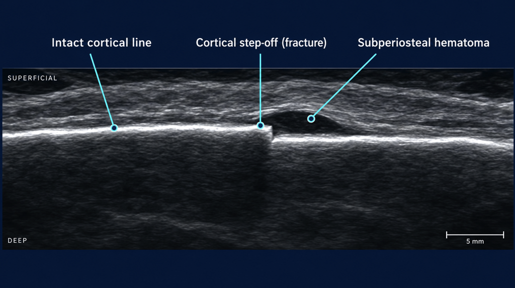
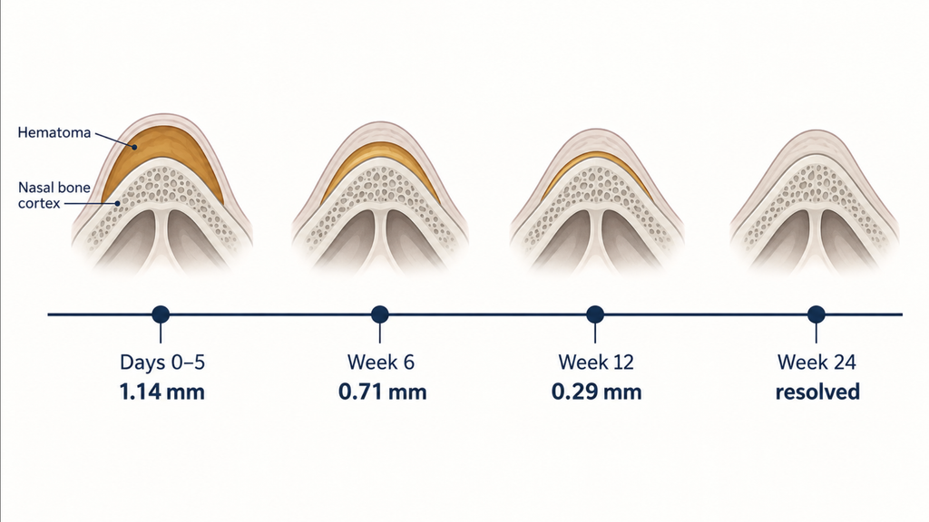
The second finding is more interesting. A six-month follow-up tracked 36 patients, all with clinically diagnosed acute fractures, so it measures timing rather than diagnostic accuracy. Subperiosteal hematoma was present in every case in the first few days after trauma, with a mean thickness of 1.14 mm. It had thinned to 0.71 mm by week six and resolved in all patients by week 24 [Nemati S, Ultrasonography Findings in Nasal Bone Fracture; 6-Month Follow-Up: Can We Estimate Time of Trauma?, 2015]. Ultrasound may therefore help distinguish a fresh fracture from an old one, which matters in medico-legal assessment.
Ultrasound for nasal bone fracture vs CT and X-ray: what the numbers say
| Study | Population | Reference | Ultrasound performance |
|---|---|---|---|
| Javadrashid, 2011 | 40 adults | CT | Sensitivity 94.9%, specificity 100% (NPV 95.3%) |
| Mohammadi, 2011 | 87 adults | Clinical exam | Sensitivity 97%, specificity 100% (CT 86%/87%; radiography 72%/73%) |
| Dogan, 2017 | 133 children, ED | Clinical exam | Radiography confirmed only 11 of 34 fractures ultrasound identified |
| Tamada, 2017 | 63 children, ED | Final diagnosis after follow-up | Sensitivity 75%, specificity 92.3% (NPV 85.7%) |
| Hassankhani, 2024 | 277 children, pooled | Varied | Pooled sensitivity 66% to 78%, specificity 87% to 88% |
Two patterns hold up. One widely repeated claim does not.
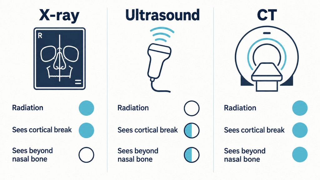
Ultrasound clearly beats plain radiography. In Mohammadi’s comparison, radiography reached 72% sensitivity and 73% specificity, well behind ultrasound [Mohammadi A, 2011]. The pediatric emergency data are starker: among 133 children with nasal trauma, radiography confirmed fractures in only 11 of the 34 cases ultrasound identified, and the authors recommended ultrasound at the bedside [Dogan S, Use of Radiography and Ultrasonography for Nasal Fracture Identification in Children Under 18 Years of Age Presenting to the ED, 2017]. Both studies used clinical examination rather than CT as the reference, so this compares bedside yield rather than formal accuracy.
Ultrasound also performs worse alone than the headline figures suggest. The two studies reporting sensitivity above 94% are both from 2011 and both small, at 40 and 87 patients. Larger, more recent pediatric work is soberer: in a prospective emergency cohort, standalone ultrasound reached only 75% sensitivity and 92.3% specificity [Tamada I, An Algorithmic Approach Using Ultrasonography in the Diagnosis of Pediatric Nasal Bone Fracture, 2017].
The claim that ultrasound matches CT rests on thin ground. Javadrashid found no significant difference between the modalities (P = 0.819) and concluded that ultrasound could serve as first-line imaging, particularly in children and pregnant patients [Javadrashid R, 2011]. That is one 40-patient study from 2011, a reasonable basis for trying ultrasound first but not for asserting equivalence.
Can ultrasound replace CT in children?
The radiation argument carries most weight here, and the evidence is at its most cautious. A 2024 systematic review pooled four studies covering 277 pediatric patients. Ultrasound showed a pooled sensitivity of 66.1% (95% CI: 35.1% to 87.5%) and specificity of 86.8% (95% CI: 80.1% to 91.4%). Excluding one outlier raised these to 78.0% and 87.8%, with a positive likelihood ratio of 6.75 and a negative likelihood ratio of 0.26 [Hassankhani A, Diagnostic Utility of Ultrasound in Pediatric Nasal Bone Fractures: A Systematic Review and Meta-Analysis, 2024].
Look at the confidence interval on that pooled sensitivity: 35.1% to 87.5%. Four studies is a thin evidence base. A positive likelihood ratio near 7 means a fracture seen on ultrasound is very likely real. A negative likelihood ratio of 0.26 lowers the probability without eliminating it.
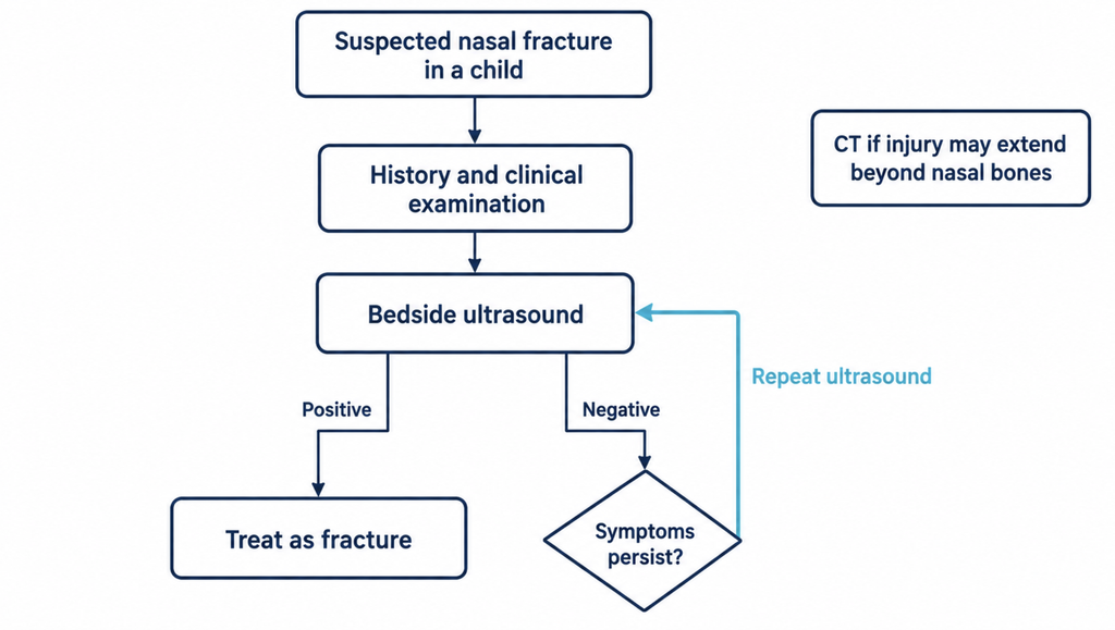
How ultrasound is deployed matters more than the pooled number. In the Tokyo cohort above, ultrasound alone reached 75% sensitivity. The management protocol it sat inside, combining history, clinical examination, and repeat scanning when symptoms persisted after a negative first scan, reached 91.7% sensitivity with a negative predictive value of 94.7%. That higher figure belongs to the whole algorithm rather than to ultrasound, and the authors were explicit that history and clinical examination remain mandatory regardless of ultrasound use [Tamada I, 2017].
So the realistic alternative to CT in children is an algorithm that uses ultrasound as one repeatable, radiation-free input, rather than a single scan.
Beyond diagnosis: guiding the reduction
Closed reduction is normally assessed indirectly, by looking and palpating. Ultrasound gives a direct view of fragment position during the procedure itself.
The available trial compared ultrasound guidance against endoscopic guidance rather than against palpation alone, so it shows how two image-guided approaches differ, not that either beats blind manual reduction. It randomized 83 children aged 12 or under with displaced nasal fractures presenting within two weeks, and severely comminuted fractures were excluded [Li C, Effect of Intraoperative Ultrasound-Guided Nasal Fracture Repair on Postoperative Child Satisfaction and Nasal Function, 2025].
The objective outcomes favored ultrasound clearly. Operative time fell from 76.8 to 57.1 minutes and hospital stay from 5.5 to 4.2 days (both P < 0.001). On rhinomanometry and acoustic rhinometry at three months, the ultrasound group had larger minimum cross-sectional area and nasal volume and lower airway resistance (all P < 0.001). Secondary repairs occurred in four control patients and none in the ultrasound group, though that comparison rests on four events and was only marginally significant (P = 0.038).
The subjective outcomes deserve more caution. Appearance and olfactory scores were rated on a visual analogue scale, and satisfaction on a questionnaire the investigators wrote themselves, completed by families with staff assistance, in a trial where neither patients nor assessors were blinded. Complication rate and overall satisfaction both favored ultrasound without reaching significance (P = 0.272 and P = 0.311). This is a single-centre study of 83 patients that has not been replicated.
The practical obstacle is acoustic coupling on an irregular curved surface. One reported workaround is a pad improvised from a medical glove [Kashimura T, Usefulness of the Glove Finger Pad for Ultrasonography in Nasal Fracture Reduction, 2024].
Where ultrasound falls short
Standalone sensitivity sits at roughly 75% in prospective pediatric emergency data, so a single negative scan does not exclude a fracture. Operator dependence is named as a limitation across this literature and is the most plausible explanation for reported sensitivity ranging from 66% to 97%.
Anything beyond the nasal pyramid is also outside its reach. Septal fractures and dislocations, orbital involvement, and wider midfacial injury fall outside what a nasal ultrasound reliably assesses. (Clinical interpretation: the studies cited here were designed around isolated nasal bone fractures and did not evaluate septal assessment.) That matters because a fractured nasal septum is a recognized predictor of closed reduction failure [Peeters N, Facial Trauma, 2016].
Heavy swelling is the last limitation, and the most common one. Marked soft-tissue edema degrades the acoustic window, which is often the state patients present in.
Clinical perspective
In clinic the useful question is which scan changes what happens next.
For most isolated nasal fractures, imaging does not decide whether to reduce. These fractures are principally diagnosed through history and clinical examination, with CT the radiographic gold standard when broader facial injury is in question [Peeters N, 2016]. Imaging documents the injury, supports planning, and reassures the patient or parent, and not all of those purposes require the highest-resolution modality available.
There is even evidence that imaging pushes decisions toward restraint. Among 117 pediatric nasal fracture patients, children who had nasal X-rays underwent closed reduction less often than those who did not (OR 0.40, 95% CI: 0.18 to 0.84, P = 0.0166), with imaging acting as a confirmatory data point that reduces unnecessary surgery in clinically false-positive cases [Leapo L, Efficacy of X-Ray in the Diagnosis of Pediatric Nasal Fracture, 2022].
Timing is the other underweighted variable. Korea’s national health information portal advises reduction within 5 to 10 days in adults and 3 to 7 days in children, before fragments begin to fix in position. An imaging study available today can be worth more than a marginally better one next week.
Operator dependence may prove solvable. A pilot deep learning algorithm trained on facial CT reached 100% sensitivity and 77% specificity against human readers, reported by its authors as a first-stage study without external validation [Jeong Y, 2024]. Extending that to point-of-care ultrasound is a plausible next step, not an established one.
Key takeaways
- Ultrasound outperforms plain radiography. In one pediatric emergency series, X-ray confirmed only 11 of 34 fractures ultrasound identified.
- As a standalone test in children, ultrasound sensitivity is roughly 66% to 78%, so a single negative scan does not exclude a fracture.
- Within a protocol using clinical examination and repeat scanning, sensitivity rose to 91.7% with a 94.7% negative predictive value.
- Subperiosteal hematoma resolves predictably over six months, which lets ultrasound help estimate when a fracture occurred.
- In one randomized trial, ultrasound-guided reduction cut operative time from 77 to 57 minutes versus endoscopic guidance, with better airway measurements at three months.
FAQ
Can ultrasound detect a broken nose?
Yes. High-resolution ultrasound visualizes cortical discontinuity in the nasal bone and the subperiosteal hematoma that accompanies a recent fracture. Reported sensitivity varies widely, and it performs best on displaced fractures of the nasal pyramid.
Is ultrasound as accurate as CT for nasal fractures?
The evidence is thinner than often claimed. A 40-patient study found no significant difference, but larger recent pediatric work reports standalone sensitivity around 75%. CT remains appropriate when injury may extend beyond the nasal bones.
Does a child need a CT for a suspected broken nose?
Not necessarily. Ultrasound is a reasonable radiation-free first step, and repeating it when symptoms persist substantially improves accuracy. A negative scan in a child with a convincing clinical picture should not end the workup. This decision belongs to the treating clinician.
Why do most hospitals still use CT or X-ray first?
Availability, operator training, and established workflow, rather than an absence of evidence. Ultrasound requires a clinician comfortable with both the probe and the anatomy, and that skill is not yet standard.
How soon should a nasal fracture be treated?
Within days rather than weeks. Korean national health guidance puts the window at 5 to 10 days for adults and 3 to 7 days for children, because fragments become harder to reposition once healing starts. Anyone with a visibly deviated nose after trauma should be assessed promptly by a clinician.
References
- Hassankhani A, Amoukhteh M, Jannatdoust P, et al. Diagnostic utility of ultrasound in pediatric nasal bone fractures: a systematic review and meta-analysis. Emerg Radiol. 2024;31(3):417-428.
- Tamada I, Mori T, Inoue N, et al. An algorithmic approach using ultrasonography in the diagnosis of pediatric nasal bone fracture. J Craniofac Surg. 2017;28(1):84-87.
- Dogan S, Kalafat UM, Yüksel B, Karaboğa T, Basturk M, Ocak T. Use of radiography and ultrasonography for nasal fracture identification in children under 18 years of age presenting to the ED. Am J Emerg Med. 2017;35(3):465-468.
- Javadrashid R, Khatoonabad M, Shams N, Esmaeili F, Jabbari Khamnei H. Comparison of ultrasonography with computed tomography in the diagnosis of nasal bone fractures. Dentomaxillofac Radiol. 2011;40(8):486-491.
- Mohammadi A, Ghasemi-Rad M. Nasal bone fracture–ultrasonography or computed tomography? Med Ultrason. 2011;13(4):292-295.
- Nemati S, Jandaghi AB, Banan R, Aghajanpour M, Kazemnezhad E. Ultrasonography findings in nasal bone fracture; 6-month follow-up: can we estimate time of trauma? Eur Arch Otorhinolaryngol. 2015;272(4):873-876.
- Li C, Li Y, Seng D, Xu Y, Ji Z. Effect of intraoperative ultrasound-guided nasal fracture repair on postoperative child satisfaction and nasal function. Lin Chuang Er Bi Yan Hou Tou Jing Wai Ke Za Zhi. 2025;39(6):559-563. Chinese.
- Kashimura T, Sugawara T, Soejima K. Usefulness of the glove finger pad for ultrasonography in nasal fracture reduction. J Craniofac Surg. 2024;35(5):e436-e438.
- Peeters N, Lemkens P, Leach R, Gemels B, Schepers S, Lemmens W. Facial trauma. B-ENT. 2016;Suppl 26(2):1-18.
- Leapo L, Uemura M, Stahl MC, Patil N, Shah J, Otteson T. Efficacy of X-ray in the diagnosis of pediatric nasal fracture. Int J Pediatr Otorhinolaryngol. 2022;162:111305.
- Jeong Y, Jeong C, Sung KY, Moon G, Lim J. Development of AI-based diagnostic algorithm for nasal bone fracture using deep learning. J Craniofac Surg. 2024;35(1):29-32.
Joonpyo Hong, MD is a board-certified otolaryngologist practicing in Korea. This article reflects his clinical interpretation of published research and does not constitute individual medical advice.
For more articles:
https://curiousmd.com/when-to-fix-a-broken-nose/
https://curiousmd.com/facial-and-neck-lacerations-by-site/
https://curiousmd.com/deviated-septum-diagnosis-ai/
Link out to:
- https://health.kdca.go.kr/healthinfo/biz/health/gnrlzHealthInfo/gnrlzHealthInfo/gnrlzHealthInfoView.do?cntnts_sn=5488 Korea Disease Control and Prevention Agency’s national health information page on nasal bone fracture. It sets out the reduction timing windows quoted above, notes that plain radiography has high false-positive and false-negative rates, and mentions intraoperative ultrasound as an option during closed reduction.
- https://www.imagegently.org/Procedures/Ultrasound The Image Gently Alliance’s guidance on ultrasound in pediatric imaging, framed around using ultrasound where possible to lower radiation dose in children. Includes plain-language material written for parents.
- https://www.choosingwisely.org/The ABIM Foundation’s Choosing Wisely campaign, which ran from 2012 to 2023 and catalogued more than 700 tests and treatments that specialty societies judged overused. Useful background for the question of whether an imaging study will change management.