
At one tertiary center, sinonasal fungus balls accounted for under 5% of surgically treated rhinosinusitis until 2001. By 2011 that share had passed 10%, and it kept climbing. The fungus did not change. The population did — older, more dental work, more CT scans finding things.
“Fungal sinusitis” sounds like one diagnosis. It is actually five, and they sit at opposite ends of a spectrum: one is a slow, harmless clump of mold that a short operation cures for good, another kills roughly half the people who get it. This article separates the five, explains who gets each one, and answers the question most readers arrive with — does this need surgery, medication, or both.
What Is Fungal Sinusitis?
Fungal sinusitis is disease of the nose and paranasal sinuses caused by fungi that have colonized or invaded them.
Fungal spores are in the air everywhere. You inhale them continuously, and they land in the nose and sinuses of healthy people every day without consequence. Disease happens only when something tips the balance: a sinus stops draining, the immune system stops policing, or the immune system overreacts. This matters for a common worry — fungal sinusitis is not usually a sign that your home has mold, and it is not contagious.
The entire classification rests on a single histologic question: has the fungus penetrated the mucosa? Everything else — prognosis, urgency, whether antifungal drugs matter — follows from that answer.
- Non-invasive: fungus sits in the sinus cavity, mucosa intact.
- Invasive: fungal hyphae cross into tissue, blood vessels, bone.
The organisms differ too. Non-invasive and chronic disease is dominated by Aspergillus species. The fulminant, life-threatening form is more often caused by Mucorales — the group behind mucormycosis.
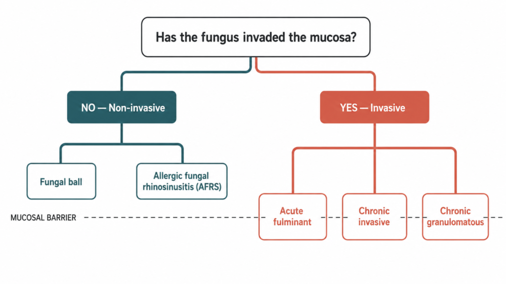
The 5 Types of Fungal Sinusitis
| Type | Invasive? | Typical patient | Time course | Core treatment |
|---|---|---|---|---|
| Fungal ball | No | Older, often female, immunocompetent | Years | Endoscopic surgery alone |
| Allergic fungal rhinosinusitis (AFRS) | No | Young, atopic, asthmatic | Months to years | Surgery + long-term steroids |
| Acute fulminant invasive | Yes | Neutropenic, uncontrolled diabetes | Days to <4 weeks | Emergency debridement + IV antifungals |
| Chronic invasive | Yes | Mildly immunocompromised, diabetic | >12 weeks | Surgery + prolonged antifungals |
| Chronic granulomatous invasive | Yes | Immunocompetent, geographically clustered | Months to years | Surgery + antifungals |
A sixth category, saprophytic fungal colonization, is sometimes listed: visible fungal growth on crusts or mucus, usually after surgery, with no tissue reaction at all. It is cleaned out during a clinic visit and rarely needs anything more.
1. Fungal ball (mycetoma)
A dense, tangled mass of fungal hyphae filling one sinus — most often the maxillary — without touching the mucosa. Patients are typically over 50, more often women, and immunologically normal. Symptoms are unimpressive: one-sided congestion, a foul smell or taste, postnasal drip, sometimes nothing at all. Many are found incidentally on a dental or brain scan.
2. Allergic fungal rhinosinusitis (AFRS)
Not an infection. An allergic reaction to fungus that is sitting harmlessly in the sinus. The immune response fills the sinuses with thick, peanut-butter-textured eosinophilic mucin and drives aggressive nasal polyps. Bent and Kuhn estimated it accounts for roughly 6% to 8% of chronic sinusitis requiring surgery. Patients are young, atopic, often asthmatic, and immunologically robust — the opposite profile from the invasive forms.
3. Acute fulminant invasive fungal sinusitis
Defined by a course of less than four weeks. Fungus invades blood vessels, causing tissue infarction and necrosis, then spreads to the orbit and skull base within days. It occurs almost exclusively in profound immunosuppression: hematologic malignancy with neutropenia, poorly controlled diabetes with ketoacidosis, transplant recipients. A pooled analysis of published series found overall survival of roughly 50%.
4. Chronic invasive fungal sinusitis
The same process running at one-hundredth the speed — more than twelve weeks, often over a year. Tissue invasion is real but slow, with dense chronic inflammation rather than infarction. It typically appears in mildly immunocompromised patients: long-standing diabetes, low-dose steroids. Because it progresses so gradually, it is frequently mistaken for a tumor or for ordinary refractory sinusitis until it reaches the orbital apex.
5. Chronic granulomatous invasive fungal sinusitis
The unusual one. Slow tissue invasion accompanied by granuloma formation and giant cells, occurring in patients with normal immune function. It is reported predominantly in Sudan, India, Pakistan, and Saudi Arabia, and is usually caused by Aspergillus flavus. Patients often present with a protruding eye or a cheek mass rather than sinus symptoms.
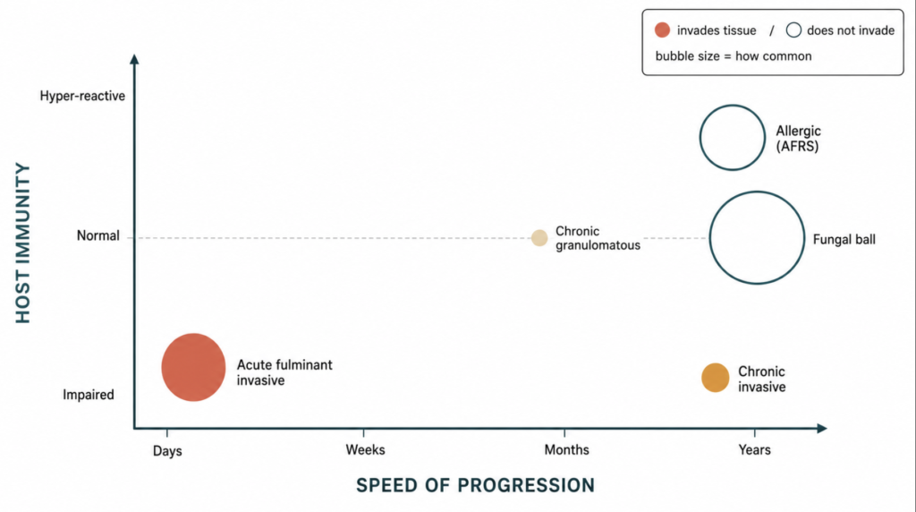
How the Five Types Differ Clinically
Four variables separate them, and each one is easy to ask about.
Speed. Acute fulminant disease evolves over days. Chronic invasive disease and fungal balls evolve over years. When a patient with a compromised immune system develops facial pain that is measurably worse than yesterday, the differential has already narrowed.
Immune status. Fungal balls and granulomatous disease occur in normal hosts. AFRS occurs in hyper-responsive hosts. Acute and chronic invasive disease occur in impaired hosts. The question “what is this patient’s neutrophil count and HbA1c?” does more diagnostic work than any single imaging finding.
Extent. A fungal ball stays inside one sinus indefinitely. AFRS expands sinuses and can thin or erode bone by pressure — bone erosion in AFRS is a mass effect, not invasion, which is why it looks alarming on CT without being an emergency. Invasive disease crosses into orbit, cavernous sinus, and brain.
Consequence. Fungal ball: cured by an outpatient operation. AFRS: controlled, often lifelong, with a high rate of revision surgery. Invasive: potentially fatal.
⚠️ Red flags — when sinus symptoms are an emergency
In anyone with uncontrolled diabetes, active chemotherapy, neutropenia, or a transplant history, the following warrant same-day evaluation:
- Facial pain out of proportion to findings, or facial numbness
- Swelling around the eye, double vision, or reduced vision
- A black or pale, insensate patch inside the nose or on the palate
- Fever with rapidly progressive one-sided sinus symptoms
Acute invasive fungal sinusitis is measured in hours to days, not weeks. Delay is the single most modifiable factor in outcome.
Who Gets Fungal Sinusitis?
Why a fungal ball forms — and who gets one
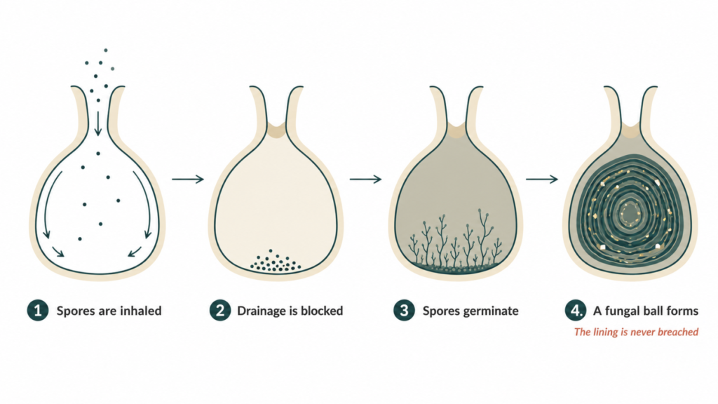
The mechanism is mechanical before it is microbial.
Fungal spores enter the sinus with every breath, and the mucociliary blanket normally sweeps them out through the natural ostium. When ventilation and drainage are impaired — by a narrow ostium, anatomic variation, chronic inflammation, or a foreign body — spores that would have been cleared instead sit in a warm, humid, low-oxygen chamber and germinate. Aspergillus tolerates low oxygen and low nutrients well, which is part of why it dominates this niche.
From there the process is additive. Hyphae accumulate into a laminated mass, calcium and heavy metal salts precipitate within it, and the result is the clay-like, gritty ball found at operation. The mucosa underneath stays intact throughout, which is why this remains a local problem no matter how long it sits there.
The dental connection is the part most patients have never heard, and it is the most practically useful.
- Endodontic treatment. Root canal sealers containing zinc oxide can extrude past the apex into the maxillary sinus. Zinc stimulates Aspergillus fumigatus growth, and endodontic treatment on maxillary teeth has been identified as a significant risk factor for maxillary fungal ball.
- Dental implants. In a series of unilateral maxillary sinus surgery, implants were more frequent in the fungus ball group, and the number of implanted teeth was significantly higher; implants penetrating more than 3 mm into the sinus were found only in the fungus ball group. The proposed mechanism is biofilm formation on the exposed implant surface.
- Periodontitis. A nationwide 12-year Korean follow-up study found periodontitis associated with subsequent development of fungal sinusitis.
Put together, the typical fungal ball patient is a woman in her sixties with normal immunity, a history of upper molar dental work, and one stubbornly blocked side.
What AFRS actually is — an allergy, not an infection
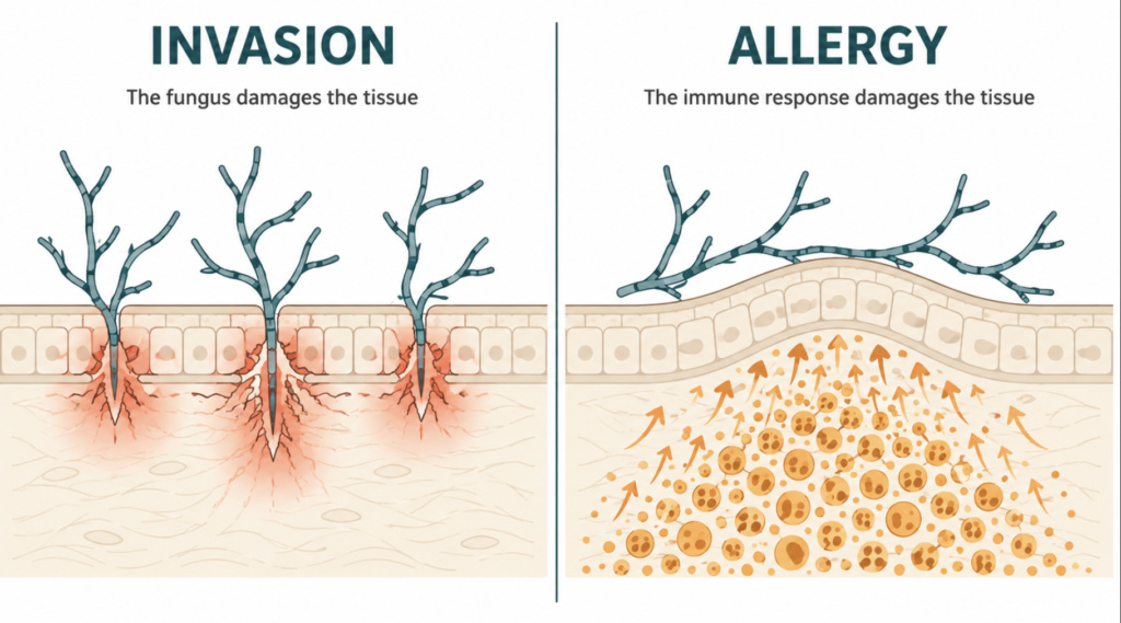
AFRS is best understood as an allergic, asthma-like reaction happening inside the sinuses.
Fungus colonizes the sinus mucus. In a susceptible person it triggers a type 2 immune response: IgE-mediated hypersensitivity to the fungus, eosinophil recruitment, and production of thick eosinophilic mucin packed with degranulated eosinophils, Charcot-Leyden crystals, and scattered fungal hyphae. The hyphae never enter the tissue. The damage comes entirely from the host’s own inflammation, which drives polyps and expands the sinus until bone remodels around it.
Bent and Kuhn’s five criteria remain the reference standard:
- Type I (IgE-mediated) hypersensitivity to fungi
- Nasal polyposis
- Characteristic CT findings
- Eosinophilic mucin without fungal invasion of tissue
- Positive fungal stain of surgically removed sinus contents
What this means about the person: AFRS signals a systemic type 2 inflammatory state. These patients usually have allergic rhinitis, frequently asthma, elevated total IgE, and peripheral eosinophilia. AFRS is a sinus manifestation of a whole-body allergic phenotype — which is why treatments developed for severe asthma have started to appear in AFRS management.
Diagnosis: How Do I Know If My Sinusitis Is Fungal?
The single most useful clue is one-sidedness.
Ordinary bacterial and inflammatory sinusitis is usually bilateral or migratory. Persistent unilateral sinus opacification is a different problem until proven otherwise — the short list is fungal ball, odontogenic sinusitis, and neoplasm. Antibiotics that fail repeatedly on one side are a signal, not a reason for another course.
CT carries most of the diagnostic weight. A fungal ball typically shows complete opacification of one sinus with internal hyperdense or frankly calcified material, plus thickening and sclerosis of the surrounding bony wall from chronic irritation. AFRS shows the opposite pattern of bone behavior: multiple sinuses filled with heterogeneous, high-attenuation mucin, with expansion and thinning or dehiscence of bone under pressure.
MRI carries a trap. Concentrated fungal material and eosinophilic mucin are so dense and dry that they lose T2 signal completely, producing a signal void that can be misread as a normally aerated sinus. An MRI reported as “clear sinuses” does not exclude fungal disease.
Endoscopy and biopsy settle it. Only histopathology distinguishes invasive from non-invasive disease — the distinction that determines whether this is an outpatient operation or a hospital admission. In a suspected acute invasive case, biopsy of suspicious mucosa is both diagnostic and urgent.
Treatment: Medication, Surgery, or Both?
The answer differs sharply by type, which is precisely why classification matters.
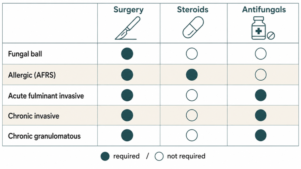
Fungal ball — surgery alone. Endoscopic sinus surgery to remove the mass and widen the sinus opening is definitive, and antifungal drugs add nothing. The fungus is not in the tissue, so a systemic drug has no vascularized target to reach. Prescribing one exposes the patient to hepatotoxicity and drug interactions for no benefit. Results are excellent, with recurrence uncommon after adequate removal.
AFRS — surgery plus sustained medical therapy. Surgery removes the polyps and every trace of eosinophilic mucin and opens the sinuses widely, but surgery alone reliably relapses. Post-operative corticosteroids — topical irrigations, with systemic courses in more severe disease — are what maintain remission. Antifungal drugs, topical or oral, are not recommended as routine therapy. For patients who relapse despite good surgery and adherent topical therapy, biologics targeting type 2 inflammation (dupilumab, omalizumab, mepolizumab) are used as a steroid-sparing option, extrapolated largely from trials in chronic rhinosinusitis with nasal polyps.
Acute fulminant invasive — three things at once, immediately. Surgical debridement of all necrotic tissue, systemic antifungal therapy, and reversal of the underlying immune defect. None of the three works without the other two, and the most important variable in survival is often how quickly neutropenia resolves or glucose control is regained.
Chronic invasive and granulomatous — surgery plus prolonged antifungals. Complete surgical removal followed by extended systemic antifungal therapy, often for months, with long-term surveillance for recurrence.
The incidental, asymptomatic fungal ball sits in a real gray zone. It is not dangerous today, and some are followed rather than operated. But a health-screening cohort found that maxillary fungus balls enlarge over time, with the observation interval the only significant predictor of growth. That argues for a considered conversation rather than automatic reassurance.
Prognosis, Recurrence, and Prevention
Outcomes track the type. A fungal ball removed completely is, for practical purposes, cured. AFRS is a chronic condition managed like severe asthma — good control is achievable, but revision surgery is needed more often than in other forms of chronic rhinosinusitis. Invasive disease carries real mortality, driven substantially by the underlying illness.
Prevention is limited but not zero. For fungal balls, the actionable item is dental: one-sided congestion or a foul smell developing in the months after a root canal, implant, or sinus bone graft on an upper molar is worth imaging rather than another antibiotic course. For invasive disease, prevention means glycemic control and a low threshold for urgent ENT evaluation during periods of neutropenia.
And to answer the question people are often too embarrassed to ask: none of these are contagious.
Clinical Perspective
Two observations shape how this diagnosis tends to play out.
The first is a threshold for imaging. Unilateral sinus symptoms that have survived two or three courses of antibiotics have crossed a line. At that point the probability of ordinary bacterial sinusitis has fallen far enough that a CT stops being defensive medicine and becomes the cheapest way to avoid a year of the wrong treatment. Fungal balls commonly reach the operating room considerably later than that threshold would suggest.
The second concerns the word itself. “Fungus in the sinus” tends to land somewhere between horror and shame — patients assume contamination, poor hygiene, a moldy home, or an immune failure nobody told them about. The reality for the most common form is a healthy woman in her sixties whose dentist did competent work on a molar a decade earlier, whose operation takes under an hour and requires no medication afterward. The gap between how the diagnosis sounds and what it usually means is one of the wider ones in this field, and closing it occupies much of the consultation.
The exception is the reason the five-part classification exists at all. The same two words, applied to a patient with a neutrophil count of 200, describe something entirely different — and outcomes in that setting depend on recognizing which of the five is present on day one rather than day five.
Key Takeaways
- Fungal sinusitis is not one disease but five, spanning outcomes from complete cure to roughly 50% mortality.
- The dividing line is whether fungus has invaded the mucosa; invasive versus non-invasive determines everything else.
- Fungal ball is the most common form, occurs in immunologically normal people, and is cured by endoscopic surgery without antifungal drugs.
- Allergic fungal rhinosinusitis is an allergic reaction rather than an infection, and requires surgery plus long-term corticosteroid control.
- Acute invasive fungal sinusitis occurs in immunocompromised patients and is a surgical emergency measured in days.
- Persistent one-sided sinusitis that does not respond to antibiotics warrants a CT scan rather than another prescription.
FAQ
Is fungal sinusitis contagious? No. Fungal sinusitis cannot be transmitted between people. The fungi involved are ubiquitous environmental organisms that everyone inhales daily; disease develops only when local drainage or host immunity changes. Family members of a patient with a fungal ball or AFRS are at no increased risk, and no isolation or precautions are needed.
Can fungal sinusitis be cured without surgery? Rarely. Every symptomatic form of fungal sinusitis has surgery at the center of treatment. A fungal ball cannot be dissolved by medication, AFRS requires physical removal of eosinophilic mucin before medical therapy can work, and invasive disease requires debridement of dead tissue. Simple saprophytic colonization is the one exception, cleared in clinic.
Do antifungal medications dissolve a fungal ball? No, and they are not recommended for it. A fungal ball sits in the sinus cavity without invading tissue, so a systemic antifungal has no vascularized target to reach. Prescribing one exposes the patient to liver toxicity and drug interactions without benefit. Complete endoscopic removal is the treatment, and it is usually definitive.
Can a root canal or dental implant cause a sinus fungal ball? They are recognized risk factors. Zinc oxide root canal sealer extruded into the maxillary sinus stimulates Aspergillus growth, and implants penetrating the sinus floor can provide a surface for fungal biofilm. Periodontitis has also been linked to later fungal sinusitis in nationwide Korean data. New one-sided sinus symptoms after upper molar dental work deserve imaging.
Is fungal sinusitis dangerous? Can it spread to the brain or eye? It depends entirely on the type. Non-invasive forms — fungal ball and AFRS — do not invade tissue and do not spread to the brain, though AFRS can expand and thin surrounding bone by pressure. Invasive fungal sinusitis genuinely does reach the orbit and skull base, and the acute form does so within days in immunocompromised patients.
How is fungal sinusitis different from regular bacterial sinusitis? The most useful practical difference is laterality and antibiotic response. Bacterial sinusitis usually involves multiple sinuses on both sides and improves with antibiotics. Fungal balls are almost always confined to one sinus and do not respond to any antibiotic course. CT adds the distinguishing features: internal calcification and thickened bony walls point toward a fungal ball.
Does a fungal ball come back after surgery? Usually not. In large endoscopic surgery series, recurrence after adequate removal and creation of a wide sinus opening is uncommon. Recurrence, when it happens, generally reflects incomplete removal or a persistent local factor such as an untreated dental source. This contrasts sharply with AFRS, which relapses frequently and requires ongoing medical management.
References
- Chakrabarti A, Denning DW, Ferguson BJ, et al. Fungal rhinosinusitis: a categorization and definitional schema addressing current controversies. Laryngoscope. 2009;119(9):1809-1818.
- deShazo RD, O’Brien M, Chapin K, Soto-Aguilar M, Gardner L, Swain R. A new classification and diagnostic criteria for invasive fungal sinusitis. Arch Otolaryngol Head Neck Surg. 1997;123(11):1181-1188.
- deShazo RD, Chapin K, Swain RE. Fungal sinusitis. N Engl J Med. 1997;337(4):254-259.
- Bent JP 3rd, Kuhn FA. Diagnosis of allergic fungal sinusitis. Otolaryngol Head Neck Surg. 1994;111(5):580-588.
- Dykewicz MS, Rodrigues JM, Slavin RG. Allergic fungal rhinosinusitis. J Allergy Clin Immunol. 2018;142(2):341-351.
- Turner JH, Soudry E, Nayak JV, Hwang PH. Survival outcomes in acute invasive fungal sinusitis: a systematic review and quantitative synthesis of published evidence. Laryngoscope. 2013;123(5):1112-1118.
- Nicolai P, Lombardi D, Tomenzoli D, et al. Fungus ball of the paranasal sinuses: experience in 160 patients treated with endoscopic surgery. Laryngoscope. 2009;119(11):2275-2279.
- Yoon YH, Xu J, Park SK, Heo JH, Kim YM, Rha KS. A retrospective analysis of 538 sinonasal fungus ball cases treated at a single tertiary medical center in Korea (1996-2015). Int Forum Allergy Rhinol. 2017;7(11):1070-1075.
- Kim JS, So SS, Kwon SH. The increasing incidence of paranasal sinus fungus ball: a retrospective cohort study in two hundred forty-five patients for fifteen years. Clin Otolaryngol. 2017;42(1):175-179.
- Park GY, Kim HY, Min JY, Dhong HJ, Chung SK. Endodontic treatment: a significant risk factor for the development of maxillary fungal ball. Clin Exp Otorhinolaryngol. 2010;3(3):136-140.
- Han SA, Kim S, Seo Y, Yang SK, Rhee CS, Han DH. Dental implant as a potential risk factor for maxillary sinus fungus ball. Sci Rep. 2024;14(1):2483.
- Kim MG, et al. Periodontitis is associated with the development of fungal sinusitis: a nationwide 12-year follow-up study. J Clin Periodontol. 2023;50(4):440-451.
- Han SC, Bae J, Park JA, et al. Natural course of the maxillary sinus fungus ball: results from Seoul National University Hospital Healthcare Center. Laryngoscope. 2025;135(4):1315-1320.
- Orlandi RR, Kingdom TT, Smith TL, et al. International consensus statement on allergy and rhinology: rhinosinusitis 2021. Int Forum Allergy Rhinol. 2021;11(3):213-739.
Joonpyo Hong, MD is a board-certified otolaryngologist practicing in Korea. This article reflects his clinical interpretation of published research and does not constitute individual medical advice.
For more interesting content:
https://curiousmd.com/sinus-microbiome-chronic-sinusitis/
https://curiousmd.com/anti-tslp-antibody-nasal-polyps/
https://curiousmd.com/epos-vs-aao-hns-chronic-rhinosinusitis-guidelines/
Link out to:
https://www.ncbi.nlm.nih.gov/books/NBK551496/
https://pubmed.ncbi.nlm.nih.gov/19544383/
https://bulletin.entnet.org/article/allergic-fungal-rhinosinusitis-management-from-clinic-to-operating-room/
https://www.nature.com/articles/s41598-024-52661-9