
Hand foot and mouth disease in adults is easy to miss, and this summer is offering plenty of chances to miss it. In week 30 of 2026, Korean sentinel surveillance recorded 36.1 suspected cases per 1,000 outpatient visits — roughly 33 times the figure from week 19. Among children aged 0 to 6, the rate reached 48.3 per 1,000, the highest of the year. Those numbers describe a childhood illness behaving exactly as expected.
What they do not capture is the adult sitting in a clinic room two weeks later with a sore throat, no mouth ulcers, and a scatter of painful blisters across the backs and palms of his hands — who has already been told twice that it is probably a viral rash of some kind.
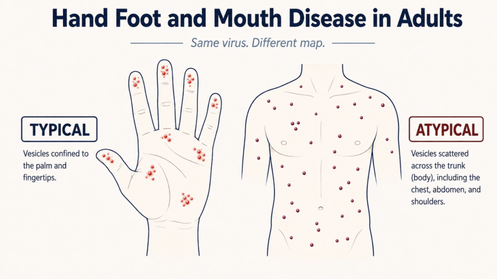
Almost everything most of us are confident about with this disease — that it belongs to children, that it comes in summer, that it appears on palms and soles — is true only of the typical case. This piece is about what happens outside it.
The Virus and What It Actually Does
Hand, foot and mouth disease (HFMD) is caused by enteroviruses, a family of small RNA viruses that enter through the mouth, replicate first in the pharynx and gut lymphoid tissue, then spread through the bloodstream to skin and mucous membranes. The blisters are not the infection itself so much as the visible endpoint of that journey [Zhu, Current Status of Hand-Foot-and-Mouth Disease, 2023].
For decades the two names that mattered were coxsackievirus(CV) A16 and enterovirus(EV)-A71. Since 2008, a third has rewritten much of the clinical picture: coxsackievirus A6, or CV-A6. The distinction is not academic trivia. The serotype largely determines which version of this disease a person gets.
Major and Minor Symptoms
The classical presentation is compact. Fever, painful ulcers in the mouth, and a vesicular rash on the palms, soles, and often the buttocks. Around those sit the minor features: sore throat, reduced appetite, malaise, sometimes diarrhoea or vomiting.
Textbooks put the incubation period at three to six days, and public health agencies repeat it. Worth knowing that a systematic review of HFMD epidemiology across Asia found no quantitative evidence supporting the incubation period for EV-A71 at all, and reported symptomatic rates for EV-A71 and CV-A16 infection ranging from 10% to 71% across four studies — a spread wide enough that the authors judged the estimates unusable for further analysis [Koh, The Epidemiology of Hand, Foot and Mouth Disease in Asia, 2016]. Several of the numbers we quote most confidently about this disease rest on thinner ground than their repetition suggests.
When a Sore Throat Is Not a Cold
Adults with HFMD typically arrive with prodromal symptoms that are indistinguishable from an upper respiratory infection: low-grade fever, fatigue, sore throat, sometimes gastrointestinal upset. That is where the diagnostic problem begins, because a sore throat in an adult is a cold until something makes it otherwise.
A few features shift the picture. In a series of 20 adults with confirmed coxsackievirus infection, oral petechiae — pinpoint haemorrhagic spots on the palate — were present in just over half, and a strawberry tongue in a quarter [Drago, Atypical Hand, Foot, and Mouth Disease in Adults, 2017]. Neither belongs to an ordinary viral pharyngitis. Painful skin lesions appearing on the hands or feet a day or two after a sore throat point in the same direction, as does a recent household contact with a child who had the disease.
This is pattern recognition rather than a diagnostic rule — the series is small, and these findings need confirmation in larger cohorts. But a sore throat that arrives with palatal petechiae and painful acral lesions is doing something a cold does not do.
Transmission, Contagiousness, and Treatment
Enteroviruses spread by the faecal-oral route, by respiratory droplets, by direct contact with blister fluid, and by contaminated surfaces and objects. The concentration of cases in nurseries and kindergartens follows from that: young children have limited hand hygiene and abundant close contact.
The infectious period is longer and messier than most parents are told. In a prospective study of 88 children hospitalised with CV-A6 HFMD, the median duration of viral shedding from pharyngeal swabs was 16 days, and more than half remained PCR-positive past current isolation recommendations [Meng, Viral Shedding Dynamics in Coxsackievirus A6 HFMD, 2026]. Viral load peaked in the first week and declined over the following five. Two caveats: these were hospitalised children, likely at the more severe end, and PCR positivity is not the same thing as being infectious.
Still, the direction is unambiguous. The CDC’s guidance that people can transmit the virus for days to weeks after symptoms resolve, and can transmit while entirely asymptomatic, matches what the shedding data show.
Treatment is supportive. There is no specific antiviral. An inactivated EV-A71 vaccine has been licensed in China, but it does not cross-protect against the other serotypes and is not available in Korea [Saguil, Hand-Foot-and-Mouth Disease: Rapid Evidence Review, 2019]. The practical priority is hydration, because painful mouth ulcers reduce fluid intake and dehydration is by a wide margin the most common complication.
The Season — and Its Exception
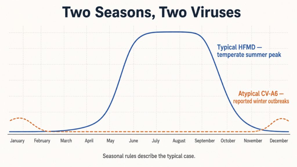
In temperate Asia, HFMD peaks in summer; in subtropical Asia, it produces spring and autumn peaks; in tropical Asia it shows no clear seasonality at all [Koh, The Epidemiology of Hand, Foot and Mouth Disease in Asia, 2016]. Korea follows the temperate pattern: cases begin rising in May and the epidemic runs through June to September.
Here is where the exception matters. Outbreaks of atypical HFMD caused by CV-A6 have been reported worldwide since 2008, and in temperate climates they have occurred predominantly in winter [Horsten, Atypical Hand, Foot, and Mouth Disease Caused by Coxsackievirus A6 in Denmark, 2018]. A Uruguayan outbreak investigation explicitly flagged winter CV-A6 activity as running counter to the summer-and-autumn expectation [Lizasoain, Hand-Foot-and-Mouth Disease in Uruguay, 2020]. These remain scattered reports rather than settled epidemiology, but the pattern is consistent enough to matter clinically.
The seasonal heuristic — it is January, so this is not HFMD — fails precisely on the cases that are hardest to recognise anyway.
Hand Foot and Mouth Disease in Adults Versus Children
Two things are true at once here, and collapsing them produces a false conclusion.
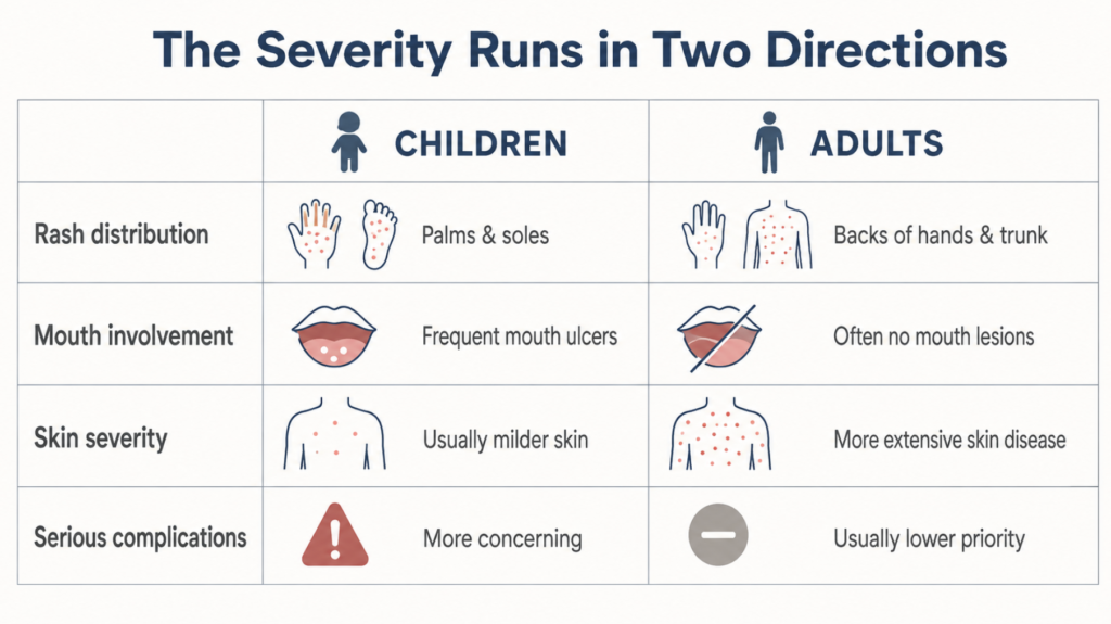
Skin disease tends to be more severe and more extensive in adults. In a retrospective series of six adults with atypical HFMD, biopsies showed massive keratinocyte necrosis and a dense cytotoxic T-cell infiltrate with strong granulysin expression — a histological picture resembling severe drug eruptions rather than a benign childhood exanthem [Second, Clinicopathologic Analysis of Atypical HFMD in Adult Patients, 2017]. Adults also show a characteristic pseudo-purpuric appearance on palms and soles, thought to reflect vesicles sitting deeper within the thicker adult palmoplantar epidermis [Neri, Hand, Foot and Mouth Disease: Clinical Features in Adult Patients, 2017].
Severe neurological and cardiopulmonary complications remain concentrated in young children, and in EV-A71. A prospective cohort study comparing serotypes found CNS complications most frequent with EV-A71, followed at some distance by CV-A4, CV-A2, CV-A10, CV-A6, and least often CV-A16 [Li, Clinical Characteristics and Severity of HFMD by Virus Serotype, 2025]. Population-based work on severe paediatric cases identified age one to three years and EV-A71 infection among the risk factors for death [Wang, Epidemiological and Clinical Characteristics of Severe HFMD Among Children, 2020].
So an adult with atypical HFMD may look far worse than the toddler who infected them while facing a considerably lower risk of the complications that actually kill. Adults are also more often asymptomatic or mildly affected — which is why household transmission from a silently infected parent is easy to miss.
One comparison worth resisting: adult case series describe lesions healing in 10 to 20 days, averaging about two weeks [Drago, Atypical Hand, Foot, and Mouth Disease in Adults, 2017], against the seven to ten days usually quoted for children. Those figures come from a small dermatology series and from public health guidance respectively, and putting them on the same axis to conclude that adults take longer overstates what the data support.
Atypical HFMD: What Gets Missed
The atypical form associated with CV-A6 breaks nearly every rule the classical description sets.
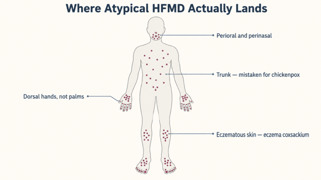
The rash goes where it should not. Vesiculobullous eruptions on the calves and dorsal hands, extensive truncal involvement, perioral and perinasal lesions, Gianotti-Crosti-like eruptions, and petechial or purpuric patterns have all been described [Chen, Arising Concerns of Atypical Manifestations in HFMD, 2023]. In one adult series, 40% had lesions confined to the extensor and lateral surfaces of the fingers, and 58% had foot lesions restricted to extensor surfaces and toe borders [Drago, Atypical Hand, Foot, and Mouth Disease in Adults, 2017]. Palms and soles — the two sites the disease is named for — can be spared.
The mouth can be clear. In the same series, erythematous-vesicular oral lesions were found in only 15% of patients, and 35% had no mucosal lesions at all. A clinician waiting for oral ulcers before considering HFMD will wait through most adult atypical cases.
It lands on damaged skin. In patients with atopic dermatitis, lesions cluster in eczematous areas, a pattern named eczema coxsackium by analogy with eczema herpeticum [Mathes, Eczema Coxsackium and Unusual Cutaneous Findings in an Enterovirus Outbreak, 2013; Horsten, Eczema Coxsackium Caused by Coxsackievirus A6, 2016].
The predictable consequence is misdiagnosis. Atypical HFMD is mistaken for varicella, eczema herpeticum, disseminated zoster, erythema multiforme major, and bullous impetigo [Chen, Arising Concerns of Atypical Manifestations in HFMD, 2023]. Truncal vesicles appeared in 95% of one adult series — enough to raise reasonable suspicion of chickenpox — though the lesions were never pustular and rarely umbilicated [Drago, Atypical Hand, Foot, and Mouth Disease in Adults, 2017]. These are small series, and the distinguishing features should be treated as clinical signposts rather than criteria.
The Complication That Arrives Late
Weeks after everything has healed, some patients watch their fingernails or toenails lift and fall off.
Onychomadesis — separation of the nail plate from the matrix — is the best-documented delayed sequela of HFMD. A meta-analysis of CV-A6 disease put the rate at 28% (95% CI 21.9–34.3%), against a severe-disease rate of only 0.10% [Zhao, Epidemiology and Clinical Presentation of Coxsackievirus A6, 2020]. Roughly one in four, with meaningful variation between studies and populations.
The timing explains why almost nobody connects the two events. In a Spanish outbreak investigation, HFMD had preceded nail shedding by an average of 40 days, with a relative risk of 14, and each affected child shed a mean of four nails, ranging from one to twelve [Guimbao, Onychomadesis Outbreak Linked to Hand, Foot, and Mouth Disease, Spain, 2010]. Adults show the same delay: in one small series, three of ten developed onychomadesis three to eight weeks after diagnosis [Neri, Hand, Foot and Mouth Disease: Clinical Features in Adult Patients, 2017]. Palmoplantar desquamation follows a similar late course.
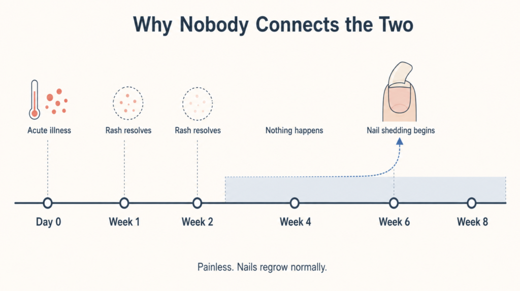
The proposed mechanism is a temporary arrest of the nail matrix during acute infection, though this remains an inference rather than a demonstrated pathway in humans. The reassuring part is not in doubt: shedding is painless, and nails regrow normally.
Clinical Perspective
Adult HFMD frequently reaches an ENT clinic before it reaches a dermatology clinic, because sore throat arrives before the rash and because the rash, when it appears, does not look like the picture in anyone’s memory. The instinct to reach for the seasonal and demographic priors — adults do not get this, it is not summer, the palms are clear — is exactly what produces the delay. Two questions cost nothing and change the pretest probability substantially: has anyone in the household had a rash illness in the last two weeks, and is there anything on the palate? Palatal petechiae in an adult with a sore throat is a finding worth pausing on.
Key Takeaways
- Hand, foot and mouth disease in adults is often asymptomatic or mild, but when it presents it frequently takes an atypical form that does not resemble the childhood illness.
- Atypical HFMD, associated with coxsackievirus A6 since 2008, can spare the palms and soles, spare the mouth entirely, and spread to the trunk, face, and areas of pre-existing eczema.
- Skin disease tends to be more severe in adults, while severe neurological and cardiopulmonary complications remain concentrated in young children and in enterovirus A71.
- Onychomadesis affects roughly one in four patients with coxsackievirus A6 disease and typically appears about five to six weeks after the acute illness — painlessly, with normal regrowth.
- Viral shedding commonly continues for around two weeks and can persist beyond standard isolation periods, so recovery from symptoms does not reliably mark the end of transmissibility.
Frequently Asked Questions
Can adults get hand, foot and mouth disease? Yes. Most adult infections are asymptomatic or mild, which is why the disease is under-recognised in adults, but symptomatic cases occur and have been reported with increasing frequency since coxsackievirus A6 emerged. Adults most often acquire it from young children in the household.
How long am I contagious? Longer than symptoms last. Viral shedding from the throat continued for a median of 16 days in one prospective study of hospitalised children, with peak viral load in the first week. PCR positivity is not identical to infectiousness, but transmission after symptom resolution — and from people with no symptoms — is well established.
Why did my fingernails fall off weeks after I recovered? This is onychomadesis, a recognised late complication of HFMD that typically appears about five to six weeks after the illness. Nail shedding is painless and nails regrow normally. The long delay is the reason most people do not connect it to the original infection.
Is it herpangina or hand, foot and mouth disease? Both are caused by enteroviruses. Herpangina produces ulcers concentrated on the soft palate and tonsillar pillars without the acral rash; HFMD combines oral lesions with skin lesions on the extremities [Saguil, Hand-Foot-and-Mouth Disease: Rapid Evidence Review, 2019]. In atypical adult cases the distinction blurs, since oral involvement may be minimal or absent.
Can you get it more than once? Yes. Immunity is largely serotype-specific, so infection with coxsackievirus A16 offers no protection against coxsackievirus A6 or enterovirus A71 [Zhu, Current Status of Hand-Foot-and-Mouth Disease, 2023]. Repeat episodes with different serotypes are entirely possible
References
Chen Y, Dai B, Han S, Duan G, Yang H, Chen S. Arising concerns of atypical manifestations in patients with hand, foot, and mouth disease. Vaccines (Basel). 2023;11(2):405.
Drago F, Ciccarese G, Broccolo F, Rebora A, Parodi A. Atypical hand, foot, and mouth disease in adults. J Am Acad Dermatol. 2017;77(2):e51-e56.
Guimbao J, Rodrigo P, Alberto MJ, Omeñaca M. Onychomadesis outbreak linked to hand, foot, and mouth disease, Spain, July 2008. Euro Surveill. 2010;15(37):19663.
Horsten HH, Fisker N, Bygum A. Eczema coxsackium caused by coxsackievirus A6. Pediatr Dermatol. 2016;33(3):e230-e231.
Horsten HH, Kemp M, Fischer TK, Lindahl KH, Bygum A. Atypical hand, foot, and mouth disease caused by coxsackievirus A6 in Denmark: a diagnostic mimicker. Acta Derm Venereol. 2018;98(3):350-354.
Koh WM, Bogich T, Siegel K, Jin J, Chong EY, Tan CY, et al. The epidemiology of hand, foot and mouth disease in Asia: a systematic review and analysis. Pediatr Infect Dis J. 2016;35(10):e285-e300.
Li Y, Yang J, Liang L, Wang K, Turtle L, Li P, et al. Clinical characteristics and severity of hand, foot, and mouth disease by virus serotype: a prospective hospital-based cohort study. PLoS Negl Trop Dis. 2025;19(5):e0013039.
Lizasoain A, Piegas S, Victoria M, Da Silva EE, Colina R. Hand-foot-and-mouth disease in Uruguay: coxsackievirus A6 identified as causative of an outbreak in a rural childcare center. J Med Virol. 2020;92(2):167-173.
Mathes EF, Oza V, Frieden IJ, Cordoro KM, Yagi S, Howard R, et al. “Eczema coxsackium” and unusual cutaneous findings in an enterovirus outbreak. Pediatrics. 2013;132(1):e149-e157.
Meng J, Cheng Y, Chen Z, et al. Viral shedding dynamics in coxsackievirus A6 hand, foot, and mouth disease: a prospective, individual-level analysis. BMC Med. 2026;24:334.
Neri I, Dondi A, Wollenberg A, Ricci L, Ricci G, Piccirilli G, et al. Hand, foot and mouth disease: an overview of clinical features in adult patients. J Eur Acad Dermatol Venereol. 2017;31(9):e386-e387.
Saguil A, Kane SF, Lauters R, Mercado MG. Hand-foot-and-mouth disease: rapid evidence review. Am Fam Physician. 2019;100(7):408-414.
Second J, Velter C, Calès S, Truchetet F, Lipsker D, Cribier B. Clinicopathologic analysis of atypical hand, foot, and mouth disease in adult patients. J Am Acad Dermatol. 2017;76(4):722-729.
Wang Y, Zhao S, Wei Y, Li K, Jiang X, Li C, et al. Epidemiological and clinical characteristics of severe hand-foot-and-mouth disease (HFMD) among children: a 6-year population-based study. BMC Public Health. 2020;20(1):801.
Zhao TS, Du J, Sun DP, Zhu QR, Chen LY, Ye C, et al. A review and meta-analysis of the epidemiology and clinical presentation of coxsackievirus A6 causing hand-foot-mouth disease in China and global implications. Rev Med Virol. 2020;30(2):e2087.
Zhu P, Ji W, Li D, Li Z, Chen Y, Dai B, et al. Current status of hand-foot-and-mouth disease. J Biomed Sci. 2023;30(1):15.
Surveillance data: Korea Disease Control and Prevention Agency, hand, foot and mouth disease sentinel surveillance, 2026.
Joonpyo Hong, MD is a board-certified otolaryngologist practicing in Korea. This article reflects his clinical interpretation of published research and does not constitute individual medical advice.
For more interesting content:
https://curiousmd.com/ramsay-hunt-syndrome-facial-paralysis/
https://curiousmd.com/shingles-vaccine-and-hearing-loss/
Link out to: